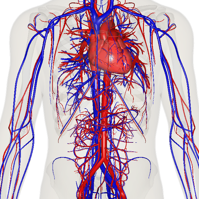
The Cardiovascular System:
Lab Objectives: The HeartAfter completing these lab exercises, you should be able to:
- Describe the location and coverings of the heart and the three layers of the heart wall.
- Identify major features of cardiac muscle tissue on a prepared microscope slide using the Gateway Virtual Microscopy software.
- Identify the major heart structures on models and images shown here and on www.essentialsofanatomyandphysiology.weebly.com.
- Describe the flow of a drop of blood through the pulmonary and systemic circulations, listing the vessels, chambers and valves.
- Describe the changes that take place in the heart after birth.
- Identify the selected heart structures on a dissected sheep heart.
- Describe each heart sound and state when each one occurs.
- Describe the relationship of ascultated heart sounds, pulse rate and heart rate.
- Identify the events responsible for systolic and diastolic pressure.
- Measure systolic and diastolic blood pressure at rest and after exercise.
- Discuss how exercise and body position affect blood pressure.
|
By BodyParts3D/Anatomography - Anatomography, CC BY-SA 2.1 jp, https://commons.wikimedia.org/w/index.php?curid=35786381
|
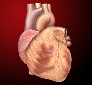
The Heart:
|
The Human Heart;By Patrick J. Lynch, medical illustrator - Patrick J. Lynch, medical illustrator, CC BY 2.5, https://commons.wikimedia.org/w/index.php?curid=1492978
|
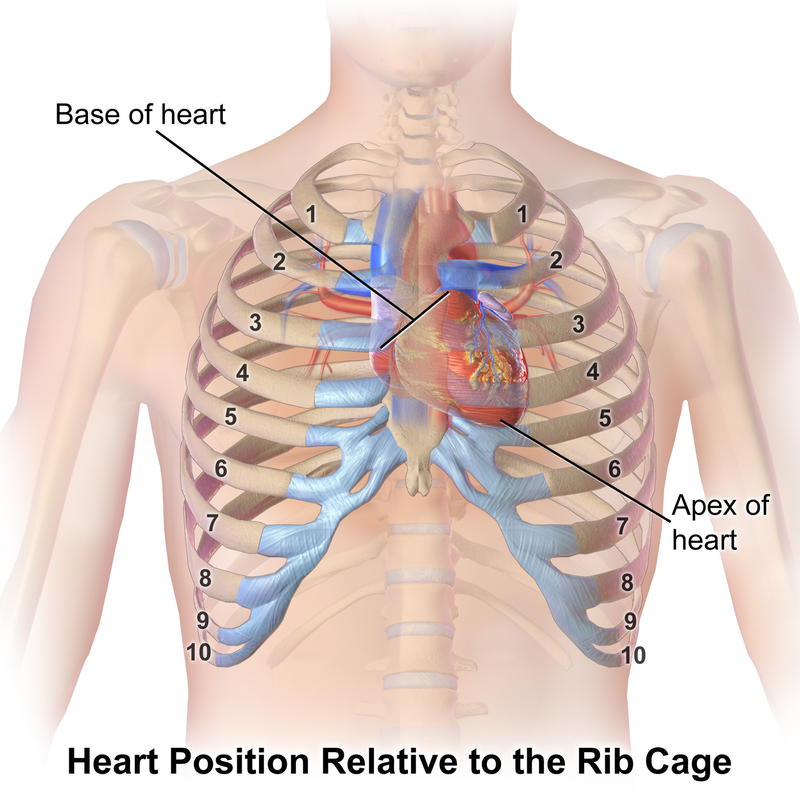
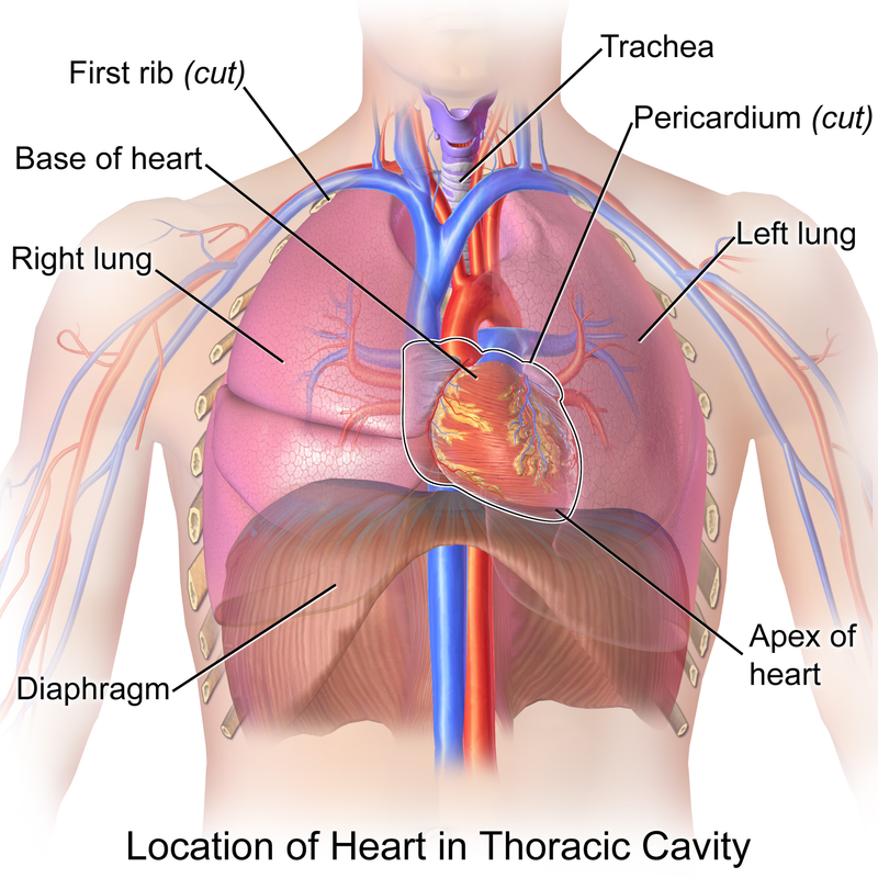
Location of the Heart:
By Blausen Medical Communications, Inc. -
|
By Blausen Medical Communications, Inc. -
|
The Pumping Mechanism of the Heart:
By DrJanaOfficial - Own work, CC BY-SA 4.0, https://commons.wikimedia.org/w/index.php?curid=50477765
Tracing the Blood Flow Through the Heart:
By Blausen Medical Communications, Inc. -
|

|
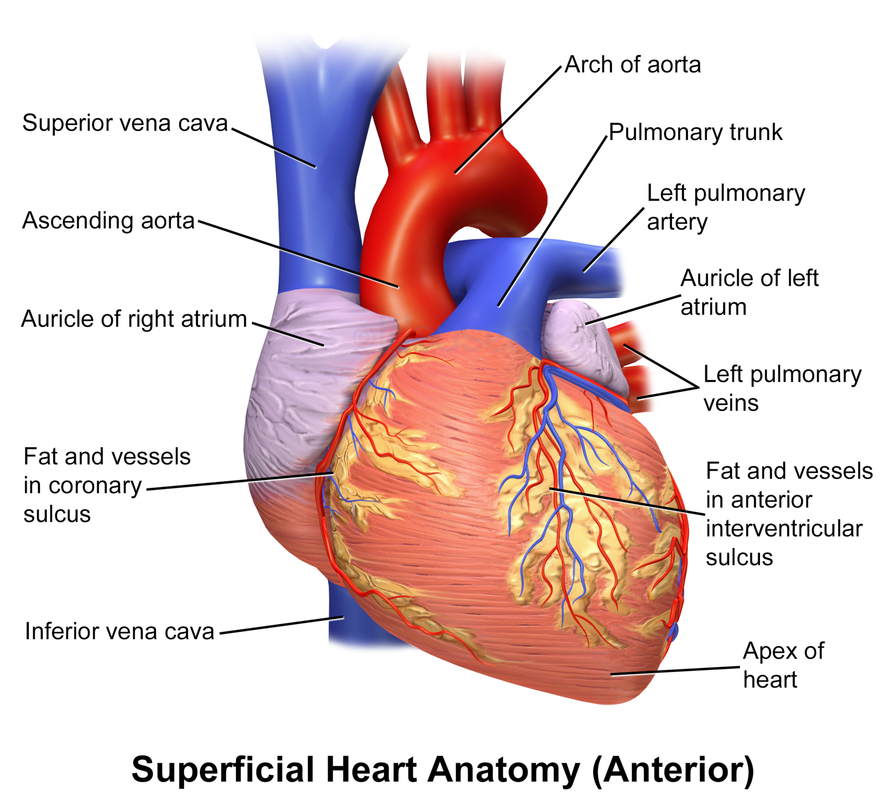
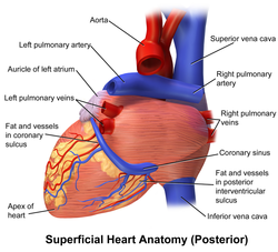
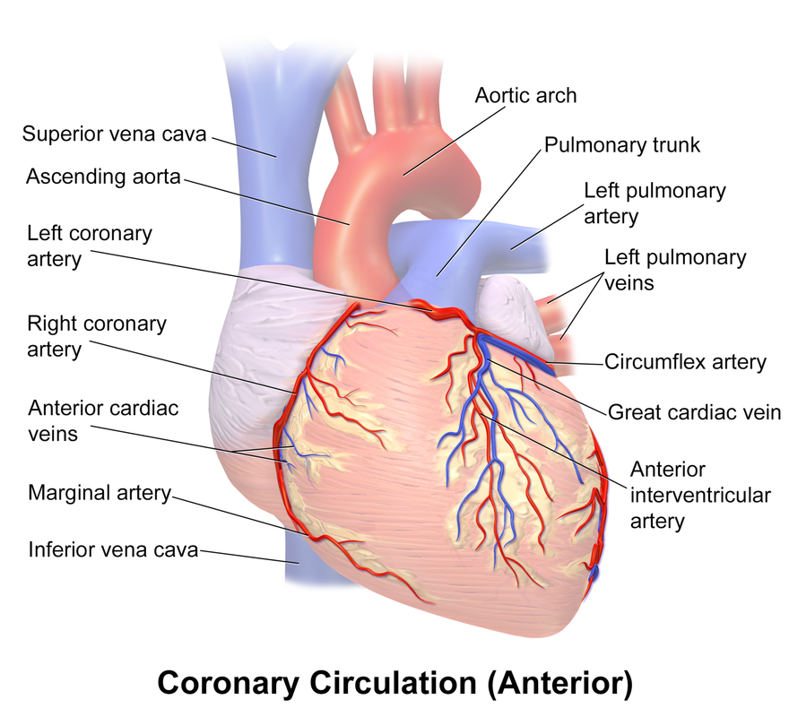
External Anatomy of the Heart:
- It is the blood supply on the heart's exterior that nourishes the myocardium of the heart
- Provided by the right and left coronary arteries, which lie in sulci, or grooves and branch out
- The coronary arteries are compressed during systole and they fill up when the heart is relaxed
- There are several cardiac veins which drain the myocardium and empty into the coronary sinus (large vein on heart posterior), which empties into the right atrium, along with a few anterior cardiac veins, which empty directly into the right atrium as well
Coverings and Layers of the Heart:
Facts about the heart:
Facts about the heart:
- It beats without external stimulation (by the involuntary, autonomic nervous system).
- It rests only between heart beats.
- It is a small double-pump that simultaneously pumps blood to and from body cells through the systemic circulation.
- It pumps blood to and from the lungs through the pulmonary circulation.
- It is about the size of your fist.
- It lies in the thoracic cavity.
- It has a main cardiac pacemaker called the sinoatrial node, but there are 2 others that can take over temporarily if it is not functioning properly
Chambers - four chambers
Sides - left and right sides
Atria - two upper chambers, forming the base of the heart
Ventricles - two lower chambers, forming the apex of the heart
Auricles - pouch-like extensions of the atria with wrinkled edges
Sulci - shallow grooves externally that mark the boundaries between the 4 heart chambers
Adipose tissue - external padding present on the heart surface
Cardiac muscle tissue - comprises the heart walls, with its own blood supply and circulation (coronary circulation)
Coronary blood vessels - encompass the heart like a crown
Right and left coronary arteries - anterior surface of the heart
Anterior interventricular branch - branch of the left coronary artery that supplies both ventricles with oxygen-rich blood and, if occluded, can result in a myocardial infarction or death.
Location of the heart:
The heart is located between the lungs in the thoracic cavity.
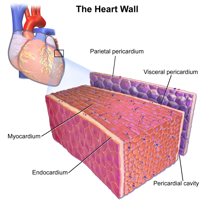
Layers of the heart:
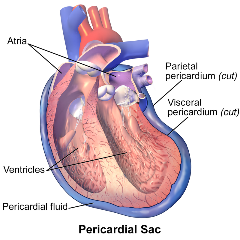
Pericardium - protects the heart and consists of an outer, tough fibrous pericardium and an inner, serous pericardium.
The wall of the heart has three layers:
The heart is located between the lungs in the thoracic cavity.
Layers of the heart:
Pericardium - protects the heart and consists of an outer, tough fibrous pericardium and an inner, serous pericardium.
- Fibrous pericardium - attaches to the diaphragm and to the great vessels of the heart.
- Serous pericardium - double-membrane composed of outer parietal layer and an inner visceral layer.
- Pericardial cavity - between the two pericardium layers; filled with serous fluid.
- Pericardial sac - the entire pericardium surrounding the heart
The wall of the heart has three layers:
- Outer epicardium - this is the visceral layer of the pericardium
- Infection is called pericarditis
- Middle myocardium - this is the cardiac muscle tissue layer, and takes up the bulk of the heart
- Infection is called myocarditis
- Inner endocardium - this is the thin layer of endothelium deep to the myocardium that lines the chambers of the heart and the valves.
- Infection is called endocarditis
- Infection is called endocarditis
By Blausen Medical Communications, Inc. - Donated via OTRS, see ticket for details, CC BY 3.0, https://commons.wikimedia.org/w/index.php?curid=26986380
|
 By BruceBlaus. When using this image in external sources it can be cited as:Blausen.com staff (2014). "Medical gallery of Blausen Medical 2014". WikiJournal of Medicine 1 (2). DOI:10.15347/wjm/2014.010. ISSN 2002-4436. - Own work, CC BY 3.0, https://commons.wikimedia.org/w/index.php?curid=30111372 By BruceBlaus. When using this image in external sources it can be cited as:Blausen.com staff (2014). "Medical gallery of Blausen Medical 2014". WikiJournal of Medicine 1 (2). DOI:10.15347/wjm/2014.010. ISSN 2002-4436. - Own work, CC BY 3.0, https://commons.wikimedia.org/w/index.php?curid=30111372
|
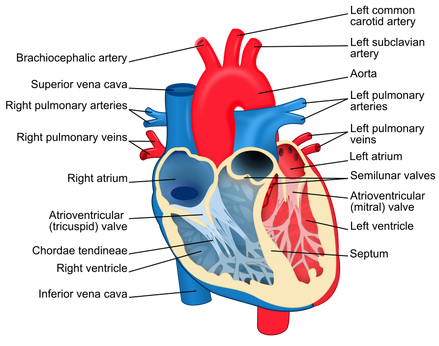
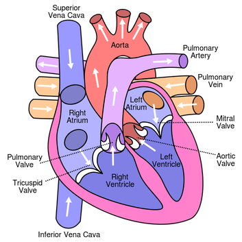
Superior vena cava - brings O2-poor blood back into the heart through the upper atria (right) from the head, neck, and arms
Inferior vena cava -brings O2-poor blood back into the heart through the upper right atria from the body inferior to the heart
Coronary sinus - returns O2-poor blood back into the heart through the upper right atria from the coronary circulation
Pulmonary trunk -blood goes from the upper right atria to the lower right ventricle then to this structure, a large artery that divides into a right and left artery that carry blood to the lungs to pick up oxygen.
Pulmonary veins - oxygen-rich blood comes back into the heart through these veins (right and left) into the left atria and ventricle.
Aorta - pumps blood to the systemic circulation and begins as a short ascending aorta, which curves to the left to form the aortic arch, descends posteriorly and continues to the descending aorta.
Ductus arteriosus - the fetal heart contains this short, temporary vessel that connects the pulmonary trunk and the aorta that acts as a right to left heart shunt that reroutes some of the blood destined for the lungs to the systemic circulation through the aorta.
Ligamentum arteriosum - ductus arteriosus changes into this after birth
By Blausen Medical Communications, Inc. - Donated via OTRS, see ticket for details, CC BY 3.0, https://commons.wikimedia.org/w/index.php?curid=26986337
Protective Coverings and Layers of the Heart:

By BruceBlaus. When using this image in external sources it can be cited as:Blausen.com staff (2014). "Medical gallery of Blausen Medical 2014". WikiJournal of Medicine 1 (2). DOI:10.15347/wjm/2014.010. ISSN 2002-4436. - Own work, CC BY 3.0, https://commons.wikimedia.org/w/index.php?curid=28761820
The Pericardium (Pericardial Sac):
By Blausen Medical Communications, Inc. - Donated via OTRS, see ticket for details, CC BY 3.0, https://commons.wikimedia.org/w/index.php?curid=26986788
- This is a double-walled serous sac surrounding the heart
- Between the double walls lies a pericardial cavity (space) filled with pericardial (serous) fluid
- The pericardial (serous) fluid reduces friction between the membranes and prevents them from "sticking" or collapsing
- Outer portion of the sac: fibrous pericardium (protection and anchoring)
- Inner portion of the sac: lies below the fibrous pericardium and includes two parts:
- Parietal pericardium-lies inside the fibrous pericardium
- Reflects back to cover the external heart at the base of the heart as visceral pericardium
- Visceral pericardium-Epicardium
- Parietal pericardium-lies inside the fibrous pericardium
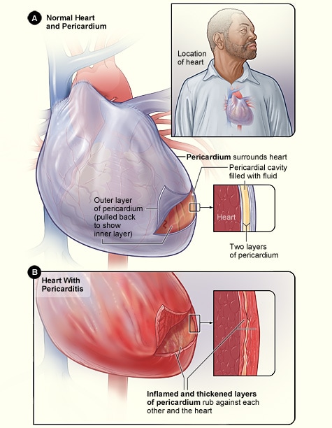
Pericarditis (Inflammation of the Pericardial Sac):
By National Heart Lung and Blood Institute (NIH) - National Heart Lung and Blood Institute (NIH), Public Domain, https://commons.wikimedia.org/w/index.php?curid=29590112
- Pericarditis is inflammation of the pericardial sac
- Causes rapid or acute onset of sharp, pleuritic chest pain under the sternum, which may radiate to the shoulders, neck or even back (bottom of scapula), along with fever, weakness, shortness of breath, sweating and heart palpitations
- Other symptoms may include dry cough, fever, fatigue and anxiety
- Symptoms are similar to myocardial infarction (heart attack) and may be misdiagnosed at first
- Pain worsens when lying down or breathing in and is often slightly better when sitting upright
- Typically caused by a viral infection like influenza, but bacteria can cause it as well
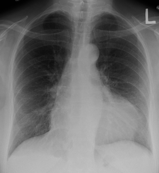
- Upon diagnosis, medical personnel will notice a pericardial rub (a friction rub heard with a stethoscope), the blood pressure may be low or lower than usual, muffled heart sounds, distention of the jugular vein, specific ECG pattern changes, and there may be fluid around the heart on the chest X-ray as seen in the images below if the condition has progressed. This is called pericardial effusion.
- Treatment includes anti-inflammatories, colchicine (possibly), steroids, aspirin, possibly antibiotics or antifungals, surgery if needed, sometimes must drain the fluid buildup
- Recovery takes a few weeks to a few months or may be chronic
- Complications: cardiac tamponade (patient will experience decreased alertness and increased lethargy), myocarditis, or constrictive pericarditis (right-sided heart failure with fluid overload, resulting in distant or muffled heart sounds)
- Affects about 3 in 10,000 people each year and those affected may have another episode and are at higher risk for
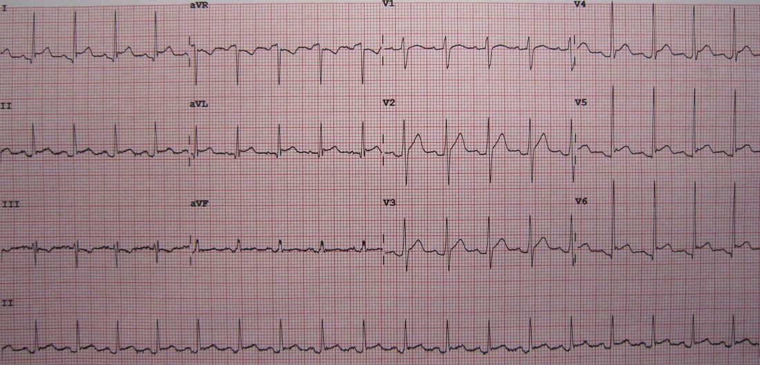
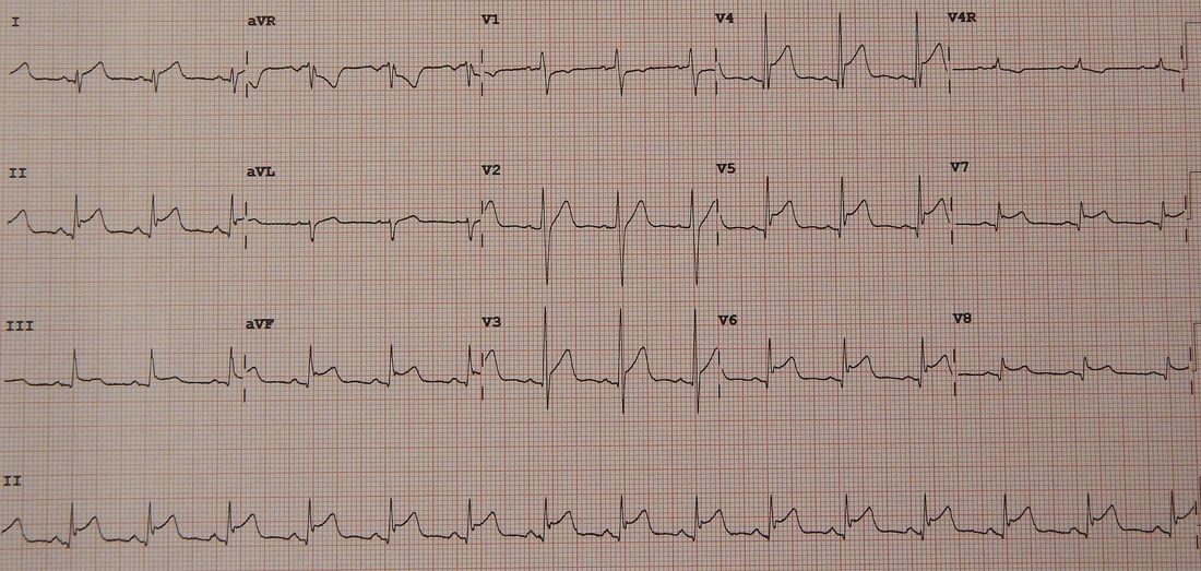
ECG Pattern in Pericarditis:
By James Heilman, MD - Own work, CC BY-SA 3.0, https://commons.wikimedia.org/w/index.php?curid=12789207
- Based on a 12-lead ECG
- ST elevations in multiple leads
- Slight reciprocal ST depression in aVR
Pericardial Effusion:
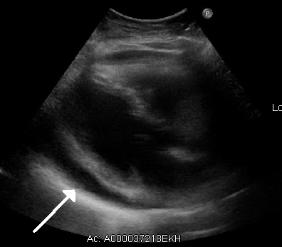
By James Heilman, MD - Own work, CC BY-SA 3.0, https://commons.wikimedia.org/w/index.php?curid=19364446
|
By James Heilman, MD - Own work, CC BY-SA 3.0, https://commons.wikimedia.org/w/index.php?curid=19364346
|
- Chest X-rays will show silhouettes indicating pericardial effusion
Infectious Pericarditis:
- Caused by a viral, bacterial or fungal infection
- Viruses: Coxsackievirus (hand-foot-mouth disease), herpesvirus, mumps virus, HIV
- Bacteria: Pneumococcus (bacterial pneumonia), mycobacterium (tuberculosis), anaerobes, streptococci (rheumatic fever)
- Fungi: Candida (yeast), Aspergillus (mold), Histoplasma (mold), Coccidioides (yeast)
- Tuberculosis is a common cause
- Idiopathic: no known cause
- Autoimmune
- Trauma
- Uremia (renal failure)
- Cancer
- Medication side effect
- Radiation
- Aortic dissection (rip in the aorta)-usually trauma causes this or an aneurysm
- Post-surgical
Clinical Lab Tests:
- Increased BUN
- If bacterial, leukocytosis (particularly neutrophils)
- If viral, leukcyotysis (particularly monocytes or lymphocytes)
- Microorganisms will show up on microscope slide and be ID'd
- Increased blood creatinine (uremic)
- Blood cultures or pericardial fluid cultures (will grow bacteria or fungi if positive; viral can be detected with PCR)
Endocardium:
- The inner lining of the heart
- Composed of simple squamous epithelium
- Covers the heart valves
- Continuous with inner lining of the great vessels
Endocarditis:

Janeway's lesions are small spots on the palms of the hands or soles of the feet, indicative of infective endocarditis;By Warfieldian - Own work, CC BY-SA 4.0, https://commons.wikimedia.org/w/index.php?curid=42835381
|
By BruceBlaus - Own work, CC BY-SA 4.0, https://commons.wikimedia.org/w/index.php?curid=57340203
|
- Endocarditis is inflammation of the endocardium of the heart, the heart's inner layer
- Inflammation may involve the heart valves (one or more of them)
- The chordae tendineae and/or inter ventricular septum are often involved as well
- Synthetic (artificial) heart valves may also become infected
- Characterized by lesions known as vegetations as seen in the image above (clumps of microorganisms, inflammatory WBCs, platelets, fibrin, and may also include a center of granulomatous tissue that may harden and calcify (fibrosis). This can permanently damage the heart (scar tissue).
- There are two types of endocarditis:
- Infective endocarditis
- Microorganism is causing the infection, usually bacteria
- Rheumatic fever can damage the heart valves and predispose the individual to infective endocarditis by placing them at higher risk for bacterial invasion. For this reason, these individuals are given prophylactic antibiotics whenever they have dental cleanings or procedures, surgeries, suspected infections or wounds to prevent this from occurring and to lower the risk.
- Rheumatic heart disease is a chronic condition caused by damaged heart valves resulting in repeated inflammation and fibrosis, leaflet valve thickening, fusion, shortening and thickening of the chordae tendineae. This can be a long-term, even severe condition.
- Noninfective endocarditis
- Cause other than a microorganism
- Often found on previously healthy, undamaged heart valves
- Vegetations that form are small, free from microbial organisms, and are typically found only along the edges or cusps of the valves
- No inflammatory response is indicated
- May be caused by a systemic (body-wide) bacterial infection, pregnancy, a venous catheter or cancer
- Risk of vegetation breaking off and causing a thrombus or embolism
- Libman-Sacks endocarditis is associated with the autoimmune disease systemic lupus erythematosus and involves the deposition of small vegetations composed of large immune complexes that have deposited on the endocardium and have provoked an immune response (inflammation)
- Infective endocarditis
- Diagnosis is based on the following tests:
- Blood cultures
- Echocardiography (cardiac ultrasound)
- ECG
- CBC with differential
- SED rate
- Signs and symptoms include cardiac murmur or palpitations, fever, chills, flu-like illness, sweating, weakness, malaise, loss of appetite, weight loss, enlarged spleen, heart failure, petechiae, Janeway's lesions
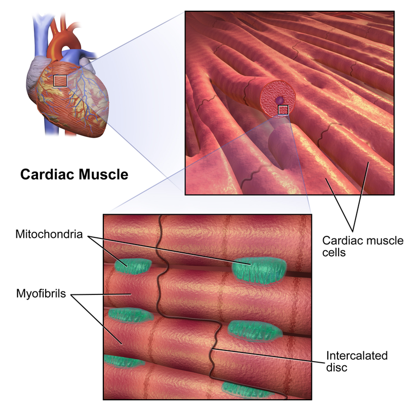
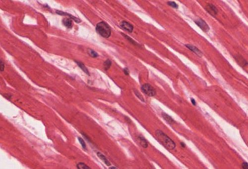
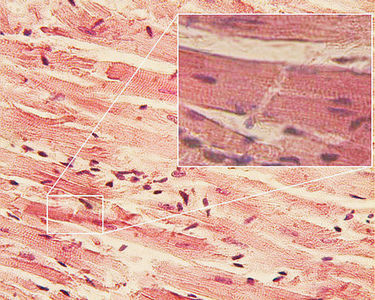
Myocardium: Middle "Muscular" Layer

|
By BruceBlaus - Own work, CC BY-SA 4.0, https://commons.wikimedia.org/w/index.php?curid=44969447
|
By OpenStax College - Anatomy & Physiology, Connexions Web site. http://cnx.org/content/col11496/1.6/, Jun 19, 2013., CC BY 3.0, https://commons.wikimedia.org/w/index.php?curid=30015036
|
By Dr. S. Girod, Anton Becker - Own work, CC BY 2.5, https://commons.wikimedia.org/w/index.php?curid=865752
|
- Muscular middle layer of the heart
- Thickest layer
- Composed mostly of cardiac muscle as seen in the images above, which is only found in the heart
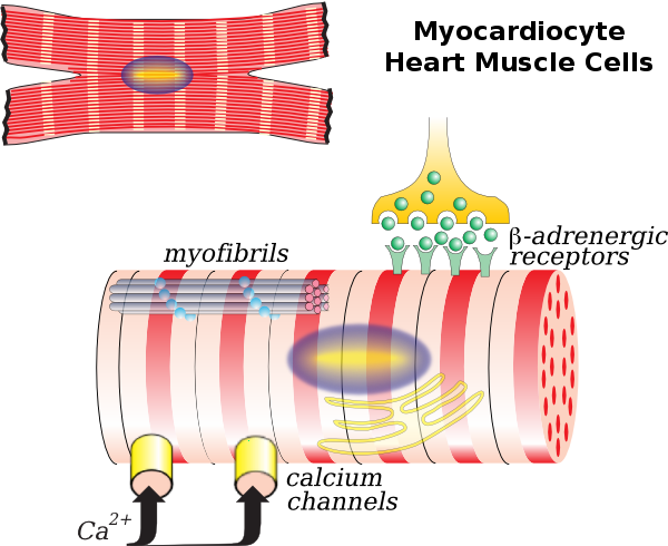
- Branched, involuntary, singular nucleus, striated, gap junctions in between cells called intercalated discs to enable coordinated heart activity and beat
- Cells are called myocytes or cardiomyocytes
- Has a spiral or figure-8 type of arrangement
Cardiomyocytes:
By OCAL (OpenClipart) - http://www.clker.com/clipart-myocardiocyte.html, CC0, https://commons.wikimedia.org/w/index.php?curid=24903488
Myocarditis:
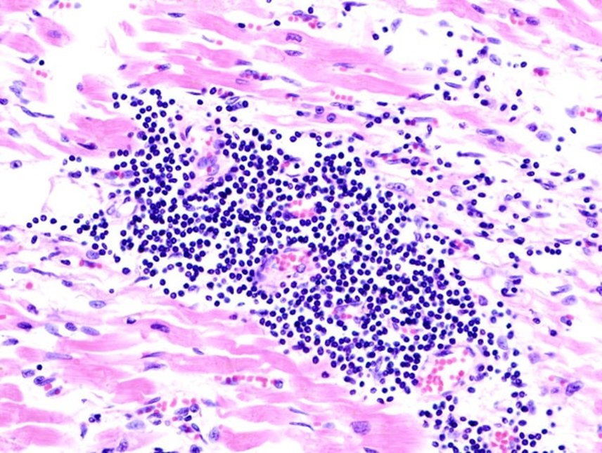
Viral Myocarditis;CC BY-SA 3.0, https://commons.wikimedia.org/w/index.php?curid=510525
By James Heilman, MD - Own work, CC BY-SA 4.0, https://commons.wikimedia.org/w/index.php?curid=49061305
- Myocarditis is inflammation of the muscular middle layer of the heart
- It is also called Inflammatory Cardiomyopthy
- Signs and symptoms: shortness of breath, stabbing, chest pain, decreased exercise tolerance, irregular heartbeat or palpitations, weakness and fatigue, congestive heart failure with swelling and edema of the limbs, puffiness, liver congestion, shortness of breath, fever, malaise, chronic cough, decreased appetite, stomach pain, symptoms resembling asthma with wheezing
- Most cases are mild
- Complications: heart failure, cardiac arrest, enlargement of the heart, sudden death (20% of all cases of sudden death in younger people)
- Usually caused by a viral infection like influenza, so the individual will typically have recently had or currently have a viral infection with fever, diarrhea, rash, pain in the joints, easily fatigue along with the symptoms listed above
- Viruses that can cause it: adenovirus (common cold, stomach bug), Parvovirus B19 (common childhood virus), Coxsackie virus (common childhood virus-hand-foot-mouth disease), Enterovirus (classic stomach bug), HIV, Rubella, Polio, Hepatitis C, Human Herpesvirus)
- Protozoans that can cause it: Trypanosoma cruzi, Toxoplasmi gondii
- The most common viral cause worldwide is Chagas Disease', carried by the "kissing bug"
- Endemic in Central and South America (infection by parasite Trypanosoma cruzi)
- These bugs are on the rise in the Southern USA
- Other causes: bacterial infection, some medications, toxins, chemicals, autoimmune disorders, pericarditis (may progress to myocarditis or occur along with it), radiation, chemotherapy, electric shock, heart attack
- Bacteria that can cause it (usually in immunocompromised individuals): streptococci, pneumococci, enterococci, staphylococci, Brucella, Corynebacterium diphtheriae, gonococcus, H. influenzae, Actinomyces, Tropheryma whipplei, Vibrio cholerae, Borrelia burgdorferi (tickborne Lyme Disease), Leptospirosis, Rickettsia, Mycoplasma pneumoniae
- Fungi that can cause it: Aspergillus
- Parasites that can cause it: Ascaris lumbricoides, Echinococcus granulosus, Paragonamus westermani, Schistosoma, Taenia solium, Trichinella spiralis, visceral larvae migrans, W. bancrofti
- Clinical lab tests for diagnosis include:
- ECG shows diffuse T-wave inversions
- Blood chemistry tests (will show increased troponin, increased creatine kinase, elevated C-reactive protein)
- CBC with differentiation (will show leukocytosis, as seen in the microscope slide above, with increase in lymphocytes and macrophages)
- ESR
- Increased IgM (Immunoserology)
- Cardiac MRI (will show inflammation)
- Heart ultrasound
- Sometimes a heart biopsy is needed with angiogram (the "gold standard" test for diagnosis)
- Treatment possibilities include antiviral medication if it is a virus, fluids, rest, ACE inhibitor, beta blocker, diuretics (to reduce edema), no exercise for awhile, corticosteroids if autoimmune, digoxin, milrinone (acute phase)
- Complications may need to be treated with implantable cardiac defibrillator or a heart transplant
- There are around 1.5 million or more cases each year in the USA alone with an average of around 300,000 deaths
- Tends to affect younger people more often than older ones
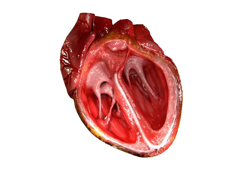
The Inner Heart Anatomy:
By ZooFari - Own workSupporting references↑ [1] (cache)↑ [2] (cache), CC BY-SA 3.0, https://commons.wikimedia.org/w/index.php?curid=9841860
|
By Blausen Medical Communications, Inc. -
|
By Own work, CC BY-SA 3.0, https://commons.wikimedia.org/w/index.php?curid=830253
|
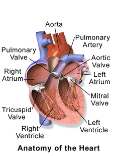
- 4 Chambers:
- 2 Superior Atria (Atrium)
- Receiving chambers
- Ineffective pumps
- Right Atrium
- Receives oxygen-poor blood from the body via the superior and inferior venae cavae (largest VEINS in the body)
- Left Atrium
- Receives oxygen-rich blood through the pulmonary veins from the lungs (only VEINS to deliver OXYGENATED blood)
- Divided by the interatrial septum
- Right Atrium
- 2 Ventricles
- Effective pumps
- Form the bulk of the heart
- Separated by interventricular septum
- Force blood to be pump out of the heart into the arteries to supply the tissues of the body or to travel to the lungs to pick up O2
- Right Ventricle
- Pumps blood into the pulmonary trunk and pushes it out to the lungs to pick up oxygen (O2)
- Left Ventricle
- Pumps blood into the large aorta and out through the arteries of the body to supply oxygen to the tissues
- Larger than the right ventricle
- Longer circuit than the pulmonary circuit above
- 2 Superior Atria (Atrium)
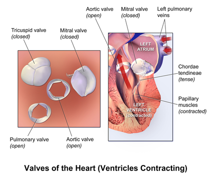
- Heart Valves
- Force one-way blood flow through the heart and prevent back flow when functioning properly
- Two Atrioventricular (AV) Valves:
- Right Atrioventricular (Tricuspid) Valve:
- Located between the right atrium and ventricle
- Prevent back flow into the atria when ventricles contract
- Has 3 cusps
- Anchored to the papillary muscles of the ventricular wall via chordae tendineae ("heart strings")
- Left Atrioventricular (Mitral or Bicuspid) Valve:
- Located between the left atrium and ventricle
- Prevent back flow into the atria when ventricles contract
- Has 2 cusps
- Anchored to the papillary muscles of the ventricular wall via chordae tendineae
- Located between the left atrium and ventricle
- Right Atrioventricular (Tricuspid) Valve:
- Pulmonary Semilunar Valve:
- Has 3 cusps
- Found between the right ventricle and pulmonary trunk
- Has 3 cusps
- Aortic Semilunar Valve:
- Has 3 cusps (pocket-like)
- Found between the left ventricle and the aorta
- Two Atrioventricular (AV) Valves:
- Force one-way blood flow through the heart and prevent back flow when functioning properly
Interior Structures of the Heart:
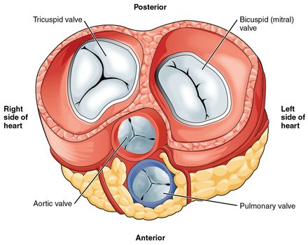
The heart has 4 valves to ensure that blood flows in one direction:
The heart has 4 valves to ensure that blood flows in one direction:
- Atrioventricular (AV) valves - two
- Right AV valve (tricuspid valve)
- Blood passing between the right atrium and right ventricle
- Left AV valve (bicuspid valve or mitral valve) - blood goes between the left atrium and left ventricle
- Chordae tendinae - string-like cords attach and secure the valves to enlarged papillary muscles, allowing the valves to close during ventricular contraction and preventing them from getting pushed up into the atria
- Papillary muscles - project from the ventricular walls
- Right AV valve (tricuspid valve)
- Semilunar valves - two
- Pulmonary Valve
- Blood in the right ventricle goes through here to enter the pulmonary trunk
- Aortic Valve
- Blood in the left ventricle goes through here to the aorta
- Pulmonary Valve
- Interventricular septum - thick wall between ventricles
- Interatrial septum - thick wall between atria
- Foramen ovale - a hole in the interatrial septum in the fetus that allows blood to bypass the lungs and go from the right atrium to the left atrium, forming another right heart to left heart shunt
- Fossa ovalis - a connective tissue membrane remnant that forms over and closes the fetal foramen after birth
By OpenStax College - Anatomy & Physiology, Connexions Web site. http://cnx.org/content/col11496/1.6/, Jun 19, 2013., CC BY 3.0, https://commons.wikimedia.org/w/index.php?curid=30148207
|
By BruceBlaus. When using this image in external sources it can be cited as:Blausen.com staff (2014). "Medical gallery of Blausen Medical 2014". WikiJournal of Medicine 1 (2). DOI:10.15347/wjm/2014.010. ISSN 2002-4436. - Own work, CC BY 3.0, https://commons.wikimedia.org/w/index.php?curid=30111374
|
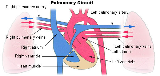
Pulmonary Circuit:
- Since the heart acts as a double-pump, the right side is the pulmonary circulation pump
- Sends CO2-rich blood to the lungs to unload CO2 and exchange it for O2 at the alveoli capillaries
- Brings blood back to the heart from the lungs into the left side of the heart (now oxygen-rich)
Public Domain, https://commons.wikimedia.org/w/index.php?curid=789677
Systemic Circuit:
- The left side of the heart carries oxygen-rich blood from left heart through the rest of the body tissues and back again to the right heart
HEART CONDITIONS: Pathophysiology
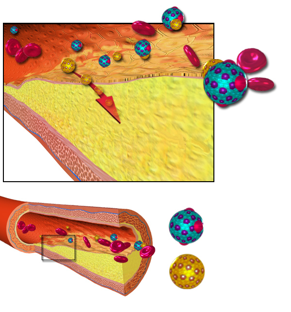
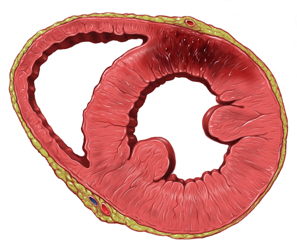
Coronary Artery Disease/Atherosclerosis:
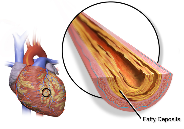
By BruceBlaus. When using this image in external sources it can be cited as:Blausen.com staff (2014). "Medical gallery of Blausen Medical 2014". WikiJournal of Medicine 1 (2). DOI:10.15347/wjm/2014.010. ISSN 2002-4436. - Own work, CC BY 3.0, https://commons.wikimedia.org/w/index.php?curid=29738538
|
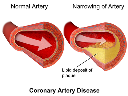
By BruceBlaus. When using this image in external sources it can be cited as:Blausen.com staff (2014). "Medical gallery of Blausen Medical 2014". WikiJournal of Medicine 1 (2). DOI:10.15347/wjm/2014.010. ISSN 2002-4436. - Own work, CC BY 3.0, https://commons.wikimedia.org/w/index.php?curid=29140355
|
Arteriosclerosis: Brought on By Atherosclerosis (Hardening of Arterial Walls and Loss of Elasticity and Flexibility
By BruceBlaus - Own work, CC BY 3.0, https://commons.wikimedia.org/w/index.php?curid=28761812
Angina: Chest Pain Due to Ischemic Condition of Vessels of the Heart That Comes and Goes and May be Stable or Unstable
By BruceBlaus. When using this image in external sources it can be cited as:Blausen.com staff (2014). "Medical gallery of Blausen Medical 2014". WikiJournal of Medicine 1 (2). DOI:10.15347/wjm/2014.010. ISSN 2002-4436. - Own work, CC BY 3.0, https://commons.wikimedia.org/w/index.php?curid=33041222
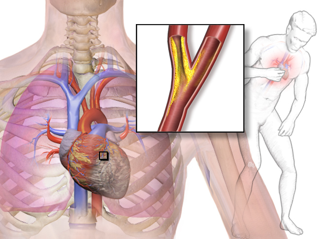
Myocardial Infarction: Heart Attack
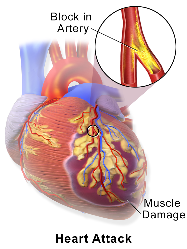
By Blausen Medical Communications, Inc. - Donated via OTRS, see ticket for details, CC BY 3.0, https://commons.wikimedia.org/w/index.php?curid=26986463
|
By Patrick J. Lynch, medical illustrator - Patrick J. Lynch, medical illustrator, CC BY 2.5, https://commons.wikimedia.org/w/index.php?curid=1490490
|
Congestive Heart Failure:
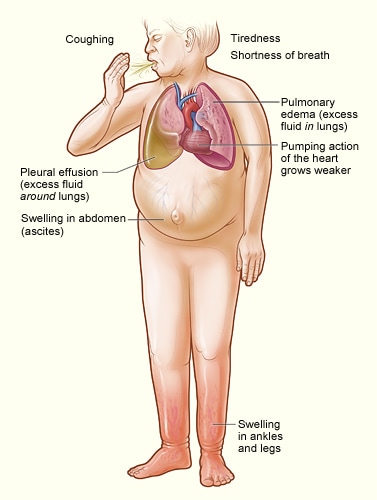
By National Heart, Lung, and Blood Institute, National Institutes of Health; originally uploaded by Wouterstomp at en.wikipedia. - http://www.nhlbi.nih.gov/health/dci/Diseases/Hf/HF_SignsAndSymptoms.html; transferred from en.wikipedia to Commons by Stevenfruitsmaak using CommonsHelper., Public Domain, https://commons.wikimedia.org/w/index.php?curid=4848343
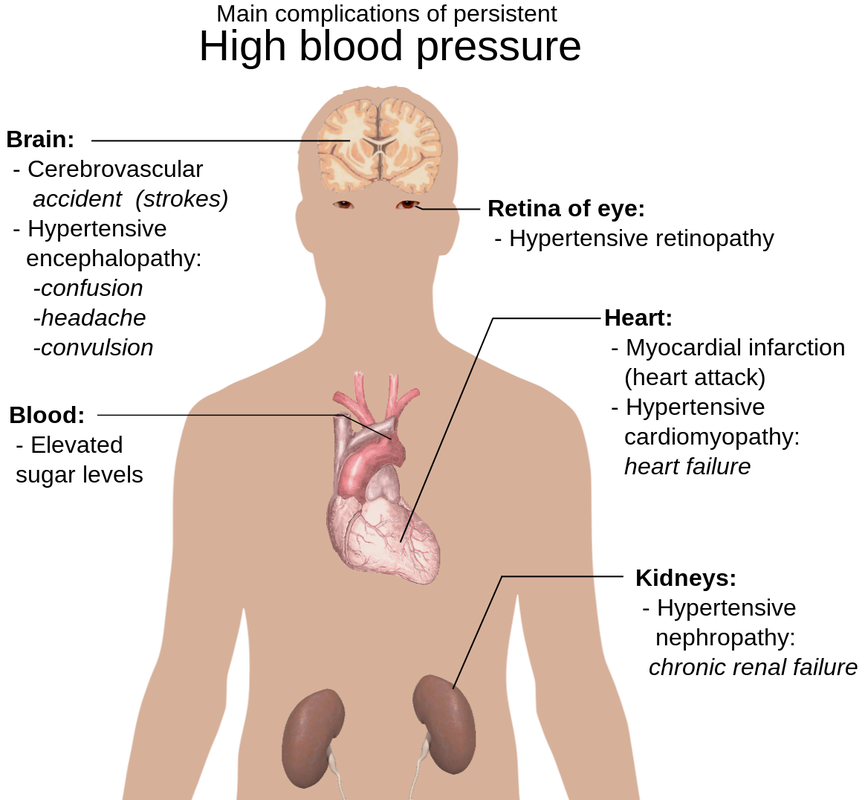
High Blood Pressure:
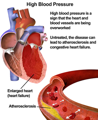
By BruceBlaus. When using this image in external sources it can be cited as:Blausen.com staff (2014). "Medical gallery of Blausen Medical 2014". WikiJournal of Medicine 1 (2). DOI:10.15347/wjm/2014.010. ISSN 2002-4436. - Own work, CC BY 3.0, https://commons.wikimedia.org/w/index.php?curid=30634277
|
By Mikael Häggström.When using this image in external works, it may be cited as:Häggström, Mikael (2014). "Medical gallery of Mikael Häggström 2014". WikiJournal of Medicine 1 (2). DOI:10.15347/wjm/2014.008. ISSN 2002-4436. Public Domain.orBy Mikael Häggström, used with permission. - All used images are in public domain., Public Domain, https://commons.wikimedia.org/w/index.php?curid=5678966
|
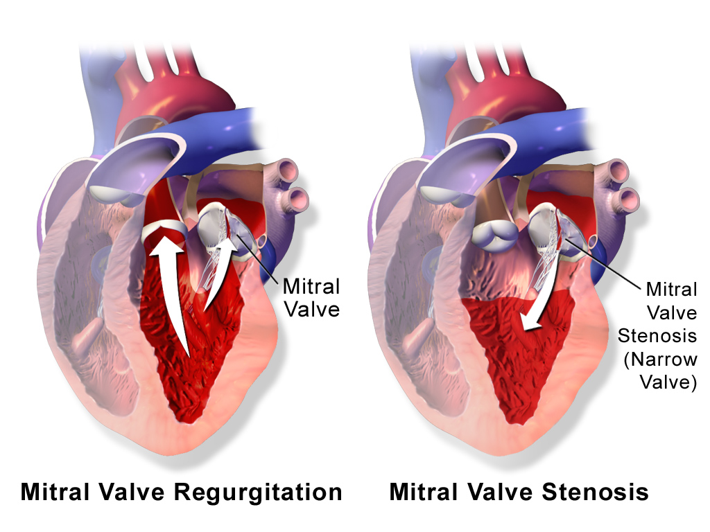
Mitral Valve Prolapse With Regurgitation and Mitral Valve Stenosis (Narrowing):
By BruceBlaus. When using this image in external sources it can be cited as:Blausen.com staff (2014). "Medical gallery of Blausen Medical 2014". WikiJournal of Medicine 1 (2). DOI:10.15347/wjm/2014.010. ISSN 2002-4436. - Own work, CC BY 3.0, https://commons.wikimedia.org/w/index.php?curid=33041242
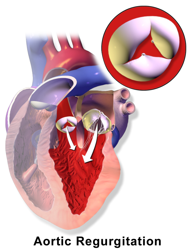
Aortic Regurgitation:
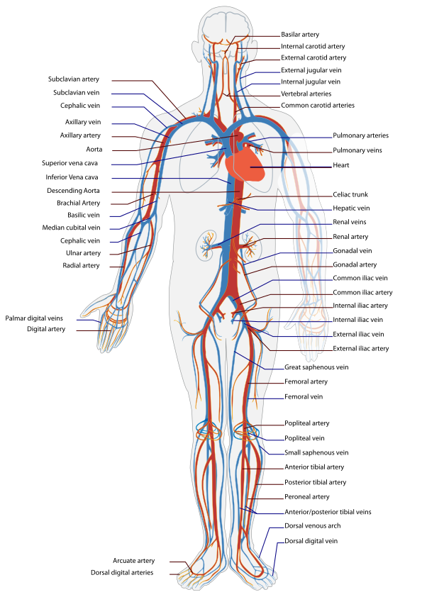
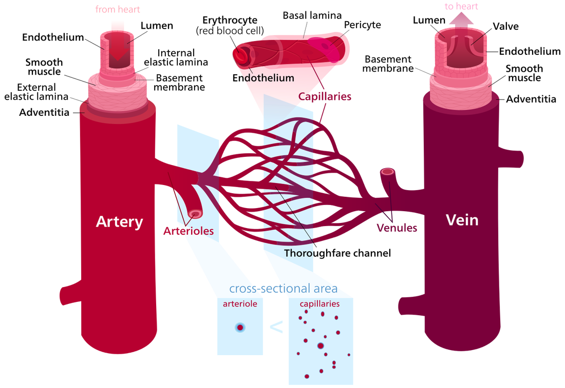
THE BLOOD VESSELS:
By LadyofHats, Mariana Ruiz Villarreal - Did myself based in the information and diagrams found in:"gray's anatomy" thirty sixth edition by Williams & Warwick."Sobotta Atlas der Anatomie des menschen" volume 1 and 2 18.Auflage by Urban & Schwarzenberg"Atlas fotografico de anatomia del cuerpo humano" 3a edicion by Yokochi, Rohen & Weinrebmultiple websites included:[1], [2], [3], [4], [5], [6], and others.This file has Outlined text. if you seek an Editable text version please use File:Circulatory System en edited.svg. or open one of the older versions of this file (down on the file history)-LadyofHats (talk) 21:42, 30 March 2010 (UTC), Public Domain, https://commons.wikimedia.org/w/index.php?curid=6698231
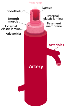
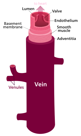
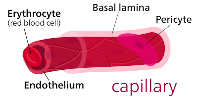
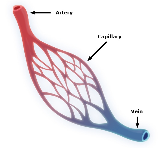
The Structure of Arteries, Veins, Capillaries:
|
|
|
|
|
|
Arteries:
|
|
Veins:
|
Capillaries:
|
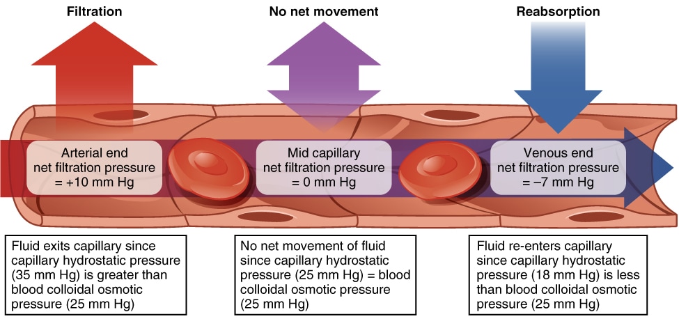
|
Heart Sounds, Pulse Rate, and Blood Pressure: Exercise 22:Auscultation - heart sounds; listening to body sounds using a stethoscope
Lubb - first sound heart during a heartbeat with the stethoscope, which is a little longer and louder thanthe second sound due to blood turbulence that occurs when the first two AV valves close during ventricular systole (top number of blood pressure)
Dupp or dubb - the second sound that occurs shortly after the first, which occurs when the ventricles relax and the two semilunar valves close, also known as diastole (bottom number of blood pressure)
Heartbeat - one cardiac cycle of lubb-dupp, pause...lubb-dupp, pause...
Heart rate- the number of heartbeats per minute; will be very close to, but necessarily equal to, the pulse or number of pulses per minute
Pulse - the blood pressure wave that is produced that travels in the arteries and can be felt when the ventricles contract
Complete Activity 1: Heart Sounds, Heart Rate, and Pulse Rate, and Discussion Questions
Blood Pressure - the pressure exerted by blood against blood vessel walls; highest in the aorta and large elastic arteries and decreases as the arteries branch and blood travels further from the heart.
Blood Pressure Gradient - the difference in blood pressure between two areas of the circulatory system.
Systolic Blood Pressure - blood pressure during ventricular systole
Diastolic Blood Pressure - blood pressure during ventricular diastole
Arterial Blood Pressure - reported in millimeters of mercury (mm Hg)
Sphygmomanometer - blood pressure cuff; used to measure systolic and diastolic blood pressure in any large artery, mainly the brachial artery
Stethoscope - placed over the brachial artery in the antecubital area, and the pressure is slowly released by opening a valve
Tissue perfusion - the body maintains blood pressure to ensure adequate blood flow to body tissues
Systemic blood pressure - increases when cardiac output increases and when resistance to blood flow increases and is affected by blood viscosity, total blood vessel length, and blood vessel diameter
Sphygmomanometer with pressure gauge, valve, cuff, and pump for taking blood pressure.
Brachial artery is shown and this is the major artery typically used to record clinical blood pressure.
Blood Pressure Gradient - the difference in blood pressure between two areas of the circulatory system.
Systolic Blood Pressure - blood pressure during ventricular systole
Diastolic Blood Pressure - blood pressure during ventricular diastole
Arterial Blood Pressure - reported in millimeters of mercury (mm Hg)
Sphygmomanometer - blood pressure cuff; used to measure systolic and diastolic blood pressure in any large artery, mainly the brachial artery
- Contains a pressure gauge attached to an inflatable rubber cuff taht is connected by a rubber tube to a hand pump (rubber bulb) or automatic pump
- It's important to use the right size cuff (pediatric, standar or large arm) to obtain an accurate reading
- Pump - used to inflate the rubber cuff to a pressure greater than the systolic pressure, which puts pressure on the artery, flattens, and stops blood flow in the artery
Stethoscope - placed over the brachial artery in the antecubital area, and the pressure is slowly released by opening a valve
- When the blood pressure is greater than the pressure in the cuff, the artery opens, blood flow returns, and the listener examines for the sound caused by the turbulent flow of blood that can be heard until blood flow returns to normal (called the Korotkoff sounds).
- Average normal systolic pressure (the first sound heard): 120 mm hG
- Average normal diastolic pressure (the last sound heard, faint): 80 mm Hg
- Written as: 120/80 mm Hg
- Venous blood pressure - average is very low (16 mm Hg)
Tissue perfusion - the body maintains blood pressure to ensure adequate blood flow to body tissues
Systemic blood pressure - increases when cardiac output increases and when resistance to blood flow increases and is affected by blood viscosity, total blood vessel length, and blood vessel diameter
- Vasodilation - increasing blood vessel diameter; decreases resistance and lowers BP
- Vasoconstriction - decreasing blood vessel diameter; increases resistance and elevates BP
Sphygmomanometer with pressure gauge, valve, cuff, and pump for taking blood pressure.
Brachial artery is shown and this is the major artery typically used to record clinical blood pressure.
Student Projects:
The Blood Vessels:
Lecture Objectives: Chapter 13, Pages 288-295Upon completion of the lectures and this chapter, you should be able to:
- Explain the relationship between blood vessel structure and function.
- Describe the parts of blood vessels.
- Differentiate between arteries, arterioles, capillaries, venules and veins.
- Trace the path of blood through the systemic, pulmonary, hepatic portal, and fetal circulations.
Lab Objectives: Exercise 23Upon completion of the lab exercises, you should be able to:
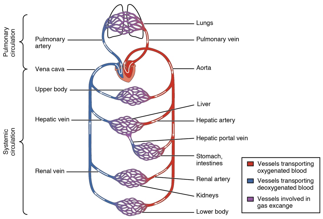
Pulmonary Circulation - takes blood from the right ventricle to the lungs and back to the heart's left atrium.
Systemic Circulation - takes blood from the left ventricle to the body tissues and back to the right atrium.
- Compare and contrast the structure of arteries, capillaries and veins.
- Identify the major arteries and veins of the systemic circulation.
- Identify the Circle of Willis and describe its function.
- Identify the major vessels of pulmonary circulation.
- Identify the major vessels of fetal circulation.
- Trace blood flow through the cardiovascular system.
Pulmonary Circulation - takes blood from the right ventricle to the lungs and back to the heart's left atrium.
Systemic Circulation - takes blood from the left ventricle to the body tissues and back to the right atrium.
Blood vessels - arteries, veins, capillaries
- Arteries - carry blood from the heart to the capillaries
- Arterioles - connect arteries to capillaries
- Veins - carry blood back to the heart
- Venules - connect veins to capillaries
- Capillaries - microscopic vessels within tissues that are thin, single-celled for O2/CO2 exchange
Blood Vessels Structure and Identification, Exercise 23*NOTE: Scroll to the images above to see lots of images of capillaries, arterioles, venules, veins and arteries and their structure.
Arteries - carry blood away from the heart and divide into smaller vessels called arterioles
Arterioles - smaller vessels that branch into tiny capillaries
Capillaries - tiny single-celled vessels for O2/CO2 exchange between blood and interstital fluid
Venules - capillaries join to form these, which merge to form larger veins
Veins - carry blood back to the heart
Systemic circulation - arteries carry oxygen-rich blood to the body tissues and veins carry oxygen-poor blood back to the heart; all major arteries of this type branch off the aorta and transport blood to different regions of the body where blood is distributed to smaller arteries and finally to arterioles that connect to capillaries.
Pulmonary circulation - arteries carry oxygen-poor blood from the right ventricle to the lungs where gas exchange occurs, and veins return oxygen-rich blood back to the left atrium of the heart
Complete Activity 1: Structure of the Blood Vessels and Activity 2: Major Arteries of the Ascending Aorta and Aortic Arch and Activity 3: Major Arteries Supplying the Head
Arteries - carry blood away from the heart and divide into smaller vessels called arterioles
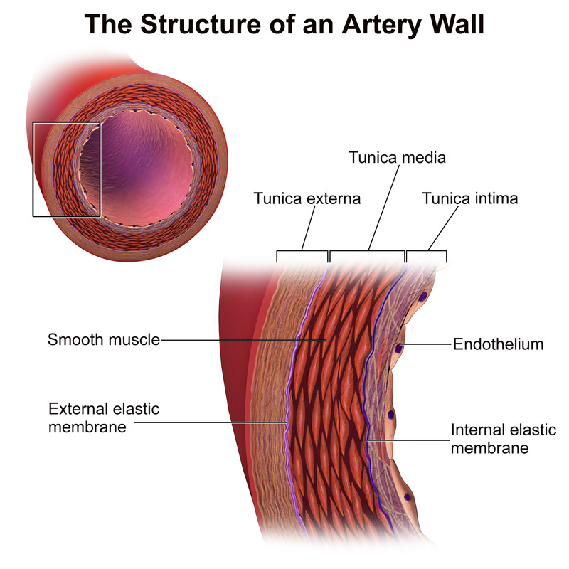
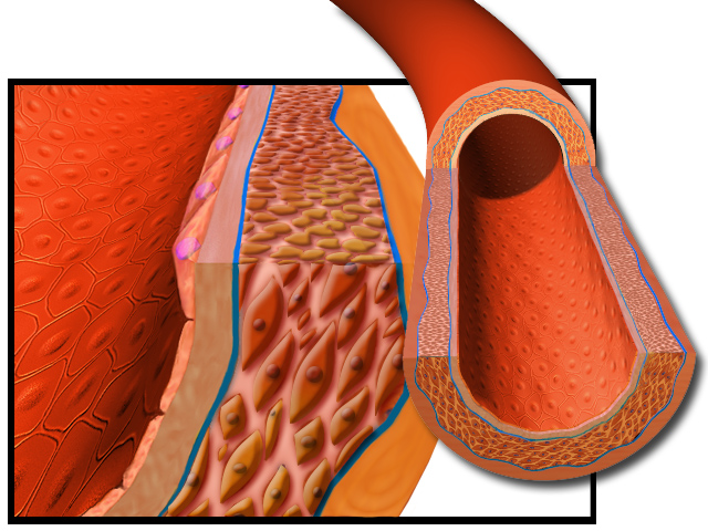
- Walls have 3 layers that enclose the center space or lumen where the blood flows
- Outer layer - elastic and collagen fibers to provide support and protection and stretch and flexibility to return to its original shape
- Middle layer - and thickest layer; contains elastic fibers and smooth muscle fibers (cells) encircling the diameter of the vessel
- Vasoconstriction - contraction of smooth muscle fibers that cause a decrease in lumen diameter
- Vasodilation - smooth muscle relaxation cause an increase in lumen diameter
- Inner layer - simple squamous epithelium (endothelium), basement membrane, and elastic tisue
Arterioles - smaller vessels that branch into tiny capillaries
- Arteries branch into these smaller and smaller little arteries from which capillaries branch
- Play a major role in controlling blood pressure and control blood flow into capillaries
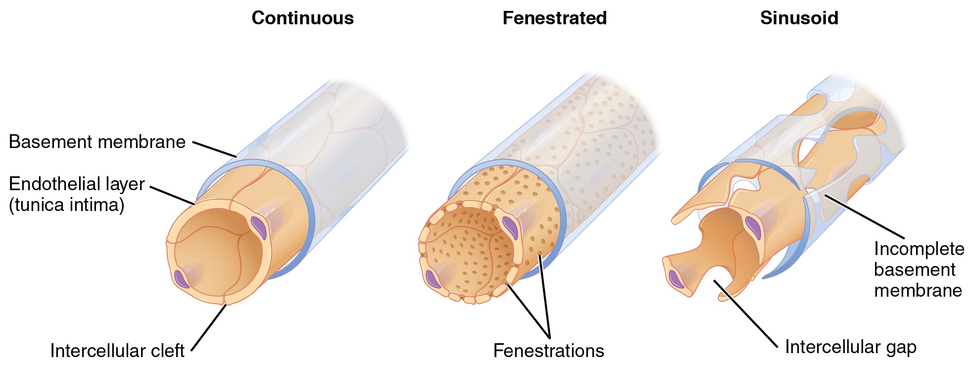
Capillaries - tiny single-celled vessels for O2/CO2 exchange between blood and interstital fluid
- Smallest diameter
- Thinnest walls
- Lumen is so small that RBC's can only pass through ONE at a time
- Single layer of endothelial cells (simple squamous epithelium)
- Basement membrane
- Exchange of substances between blood and tissues occurs ONLY here
- Structurally permeable
Venules - capillaries join to form these, which merge to form larger veins
- Blood flows from capillaries into these little veins, which drain into veins
Veins - carry blood back to the heart
- Blood flows from venules into veins
- Walls contain three layers
- Thinner walls with fewer smooth muscle fibers and elastic fibers
- Lumen of veins is much larger than arteries and often appears collapsed in tissue sections
- Contain about 60% of the total blood volume
- Blood reservoirs
- Blood pressure gradient here is very small and is often not enough to overcome gravity
- Muscular activity squeezes the veins and pushes blood toward the heart
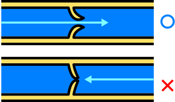
- Valves - prevent backflow of blood
Systemic circulation - arteries carry oxygen-rich blood to the body tissues and veins carry oxygen-poor blood back to the heart; all major arteries of this type branch off the aorta and transport blood to different regions of the body where blood is distributed to smaller arteries and finally to arterioles that connect to capillaries.
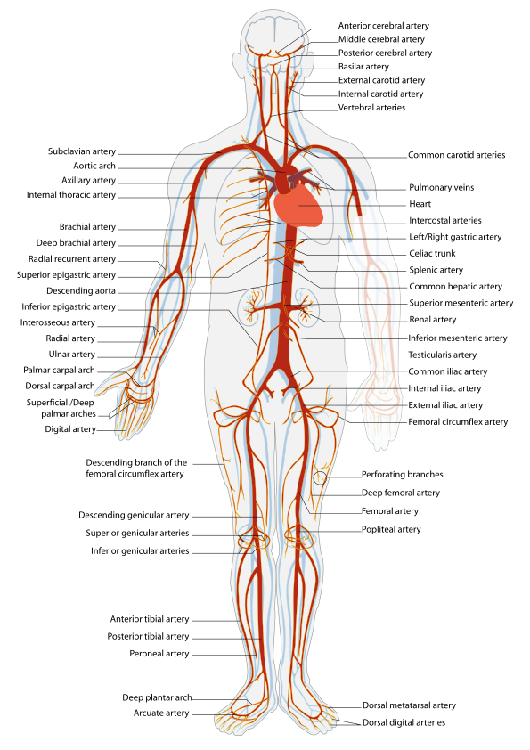
- Ascending aorta - blood is ejected from the left ventricle into this structure and it curves superiorly toward the left and becomes the aortic arch
- Right and left coronary arteries - branch off the ascending aorta near its origin and travel to the heart
- Aortic arch - three major arteries branch off of it:
- Brachiocephalic trunk - divides to form the right common carotid and the right subclavian arteries
- Left common carotid artery
- Branches to form the internal and external carotid arteries
- Internal carotid artery - travels through the neck and enters the skull through the carotid canal and supplies blood to the anterior and middle brain areas
- External carotid artery - travels along the lateral surface of the neck and terminates as two arteries near the temporomandibular joint to supply structures external to the skull.
- Branches to form the internal and external carotid arteries
- Left subclavian artery
- Vertebral arteries - two; one branches off each subclavian artery
- Travel superiorly through the transverse foramen of the cervical vertebrae and foramen magnum to supply blood to the posterior brain areas
- Vertebral arteries - two; one branches off each subclavian artery
Pulmonary circulation - arteries carry oxygen-poor blood from the right ventricle to the lungs where gas exchange occurs, and veins return oxygen-rich blood back to the left atrium of the heart
Complete Activity 1: Structure of the Blood Vessels and Activity 2: Major Arteries of the Ascending Aorta and Aortic Arch and Activity 3: Major Arteries Supplying the Head
Major Arteries of the Upper Extremities:
Axillary Artery - the subclavian artery is renamed this within the axilla (armpit)
Brachial Artery - axillary artery changes to this as it enters the arm
Radial Artery - as the brachial artery enters the forearm, it divides to form this and the ulnar artery
Superficial palmar arch - supplies blood to fingers and palms
Deep palmar arch - supplies blood to fingers and palms
Complete Activity 4: Arteries of the Upper Extremities
Major Arterial Branches of the Descending Aorta:
Thoracic aorta - lies to the left of the midline, just anterior to the vertebral column and has branches that supply thoracic structures
Abdominal aorta - anterior to the vertebral column closer to the midline, supplying the abdomen, pelvis, and lower extremities; terminates as the paired common iliac arteries
Celiac trunk - branches off the descending aorta, dividing into the left gastric artery, splenic artery, and common hepatic artery, the superior mesenteric artery, paired renal arteries, and inferior mesenteric artery
Complete Activity 5: Arterial Branches of the Descending Aorta
Major Arteries of the Pelvis and Lower Extremities:
Common iliac arteries - divide into the following:
Common iliac arteries - divide into the following:
- Internal iliac arteries - medial terminal branches off of the common iliac arteries just anterior to the lumbosacral joint, and course posteriorly to the pelvis.
- External iliac artery - is larger than the internal iliac arteries and becomes the femoral artery as it enters the thigh
Femoral artery - descends along the middle of the anterior 2/3rds of the thigh and travels to the posterior thigh, becoming the popliteal artery as it enters the posterior knee area
Popliteal artery - posterior knee; divides into the following:
Complete Activity 6: Major Arteries of the Pelvis and Lower Extremities
Popliteal artery - posterior knee; divides into the following:
- Anterior tibial artery - travels to the anterior surface of the leg; descends to the ankles, becoming the dorsalis pedis artery
- Posterior tibial artery - descends along the posterior aspect of the leg
Complete Activity 6: Major Arteries of the Pelvis and Lower Extremities
Systemic Veins:
Veins - carry blood from the capillaries back toward the heart, and are found under the skin (superficial) or deep within the body, typically near an artery.
Veins carrying blood from the lungs to the left side of the heart carry oxygen-rich blood and are part of the pulmonary circulation.
Veins carrying blood from all other body tissues carry oxygen-poor blood and are part of the systemic circulation.
Thrombus - a blood clot in a deep vein, which can cause a pulmonary embolism if not treated with an anticoagulant (deep vein thrombosis, or DVT). If it is in a superficial vein, it does not tend to form an embolus.
Embolus - a blood clot that is moving or has moved and can cause a blockage, stroke, pulmonary embolism, or even death.
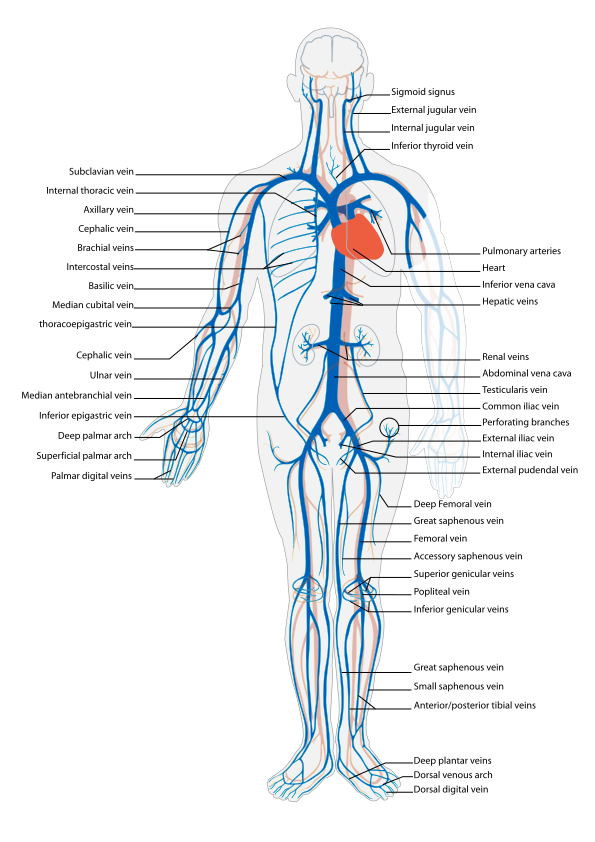
Superior vena cava - carries oxygen-poor blood into the right atrium.
Inferior vena cava - carries oxygen-poor blood into the right atrium.
Coronary sinus - carries oxygen-poor blood into the right atrium.
Complete Activity 7: Veins Carrying Blood to the Heart
Veins - carry blood from the capillaries back toward the heart, and are found under the skin (superficial) or deep within the body, typically near an artery.
Veins carrying blood from the lungs to the left side of the heart carry oxygen-rich blood and are part of the pulmonary circulation.
Veins carrying blood from all other body tissues carry oxygen-poor blood and are part of the systemic circulation.
Thrombus - a blood clot in a deep vein, which can cause a pulmonary embolism if not treated with an anticoagulant (deep vein thrombosis, or DVT). If it is in a superficial vein, it does not tend to form an embolus.
Embolus - a blood clot that is moving or has moved and can cause a blockage, stroke, pulmonary embolism, or even death.
Superior vena cava - carries oxygen-poor blood into the right atrium.
Inferior vena cava - carries oxygen-poor blood into the right atrium.
Coronary sinus - carries oxygen-poor blood into the right atrium.
Complete Activity 7: Veins Carrying Blood to the Heart
Major Veins Draining the Head and Neck:
Internal jugular veins - drain blood from the head and neck and run lateral to the internal carotid artery and common carotid artery.
External jugular veins - drain blood from the head and neck and are superficial veins descending along the lateral surface of the neck.
Vertebral veins - drain blood from the head and neck and descend through the transverse foramina of the vertebral column with the vertebral arteries.
Complete Activity 8: Major Veins Draining the Head and Neck.
Major Veins Draining the Upper Extremities:
Superficial veins - visible veins running beneath the skin of the arm, which become more visible with age.
Cephalic vein - major superficial vein of the upper extremities, which travels along each arm along the lateral surface of the anterior limb and merges with the axillary vein inferior to the clavicle.
Basilic vein - major superficial vein of the upper extremities, which travels along the medial surfaces of each posterior forearm and the medial surface of the anterior arm.
Median cubital vein - vein that is a common site for obtaining venipuncture samples; anterior to the elbow and connects the basilic and cephalic veins.
Complete Activity 9: Major Veins Draining the Upper Extremities
Major Veins Draining the Thorax, Abdomen, and Pelvis:
Internal iliac veins - veins of the pelvis that merge to form the common iliac veins
External iliac vein - veins of the pelvis that merge to form the common iliac veins
Common iliac veins - unite to form the inferior vena cava
Inferior vena cava - brings unoxygenated blood back to the right atrium of the heart
Renal veins - major veins that drain directly into the inferior cava from the kidneys
Hepatic veins - major veins that drain directly into the inferior cava from the liver.
Hepatic portal circulation - include the major veins of the hepatic circulation, which are the inferior mesenteric vein, splenic vein, superior mesenteric vein, and hepatic portal vein, which drain the stomach, intestines, spleen, pancreas, and gallbladder.
Inferior mesenteric vein - drains blood from the large intestine and joins the splenic vein.
Splenic vein - carries blood from the stomach, pancreas and spleen.
Superior mesenteric vein - drains blood from the small intestine and merges with the splenic vein to form the hepatic portal vein.
Hepatic portal vein - carries nutrient-rich blood to the liver for processing.
Azygous vein - part of the azygous system of veins that drain thoracic structures.
Complete Activity 10: Major Veins Draining the Thorax, Abdomen, and Pelvis
Major Veins Draining the Lower Extremities:
Great saphenous vein - major superficial vein of the leg; longest vein in the body and travels along the medial surface of the leg and the thigh.
Anterior tibial veins - ascend in the anterior and posterior leg, and unite inferior to the popliteal fossa to form the popliteal vein
Posterior tibial veins - ascend in the anterior and posterior leg, and unite inferior to the popliteal fossa to form the popliteal vein
Popliteal vein - ascends along the posterior surface of the knee and becomes the femoral vein that travels up the posterior thigh and becomes the external iliac vein in the pelvis
Femoral vein - deep vein that travels up the posterior thigh and becomes the external iliac vein in the pelvis
External iliac vein - deep pelvic vein
Pulmonary Circulation:
Pulmonary circulation - carries oxygen-poor blood from the right ventricle to the capillaries of the lung where oxygen is added and carbon dioxide is removed.
Pulmonary trunk - carries oxygen-poor blood from the right ventricle and divides to form right and left pulmonary arteries.
Pulmonary arteries - right and left arteries that carry blood to the lungs.
Pulmonary veins - carry oxygen-rich blood from the lungs to the left atrium.
Complete Activity 12: Major Blood Vessels of the Pulmonary Circulation
Fetal Circulation:
Placenta - exchange through the mother's circulatory system provides the fetus with oxygen and nutrients and eliminates carbon dioxide and other wastes from fetal blood here, which forms within the uterus during early pregnancy. Uterine blood vessels enter here, and substances are exchanged by diffusion between maternal and placental capillaries without direct mixing of maternal and fetal blood.
Umbilical arteries - consist of two arteries that carry oxygen-poor fetal blood to the placenta.
Umbilical vein - consists of one vein that carries oxygen-rich blood back to the fetus.
Ductus venosus - allows most of the blood to bypass the liver and enter the inferior vena cava.
Foramen ovale - allow blood to bypass the fetal lungs. After birth, vascular changes occur to allow blood to enter the lungs.
Ductus arteriosus - allow blood to bypass the fetal lungs. After birth, vascular changes occur to allow blood to enter the lungs.
Complete Activity 13: Fetal Circulation
