Bone: Osseous Tissue:
- There are approximately 206 bones in the adult body (some are or may be fused)
- There are two basic kinds of osseous tissue:
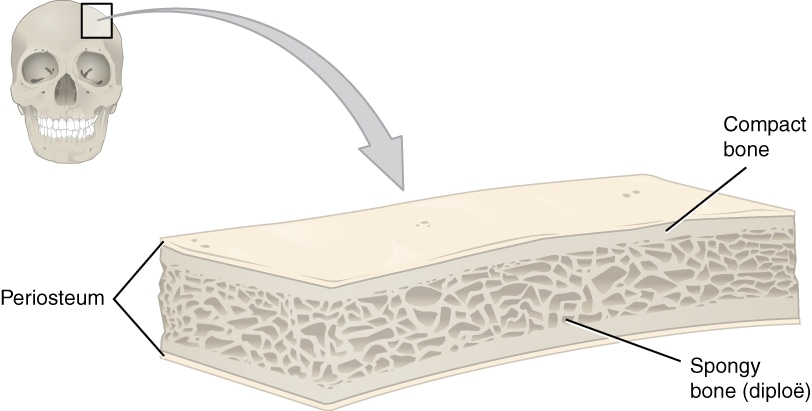
- Compact bone: smooth, dense
- Spongy bone: has lots of small airy spaces called trabeculae and looks "spongy"
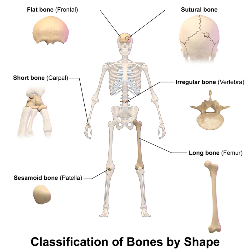
- Classification of Bones:
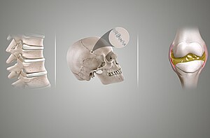
- Long Bones: mostly compact bone consisting of a shaft with heads at both ends (Examples: femur (thigh bone), humerus (arm bone), phalanges (fingers and toes))
- Humerus, radius, ulna (arms)
- Femur, tibia, fibula (legs)
- Metacarpals, phalanges (fingers)
- Metatarsals, phalanges (toes)
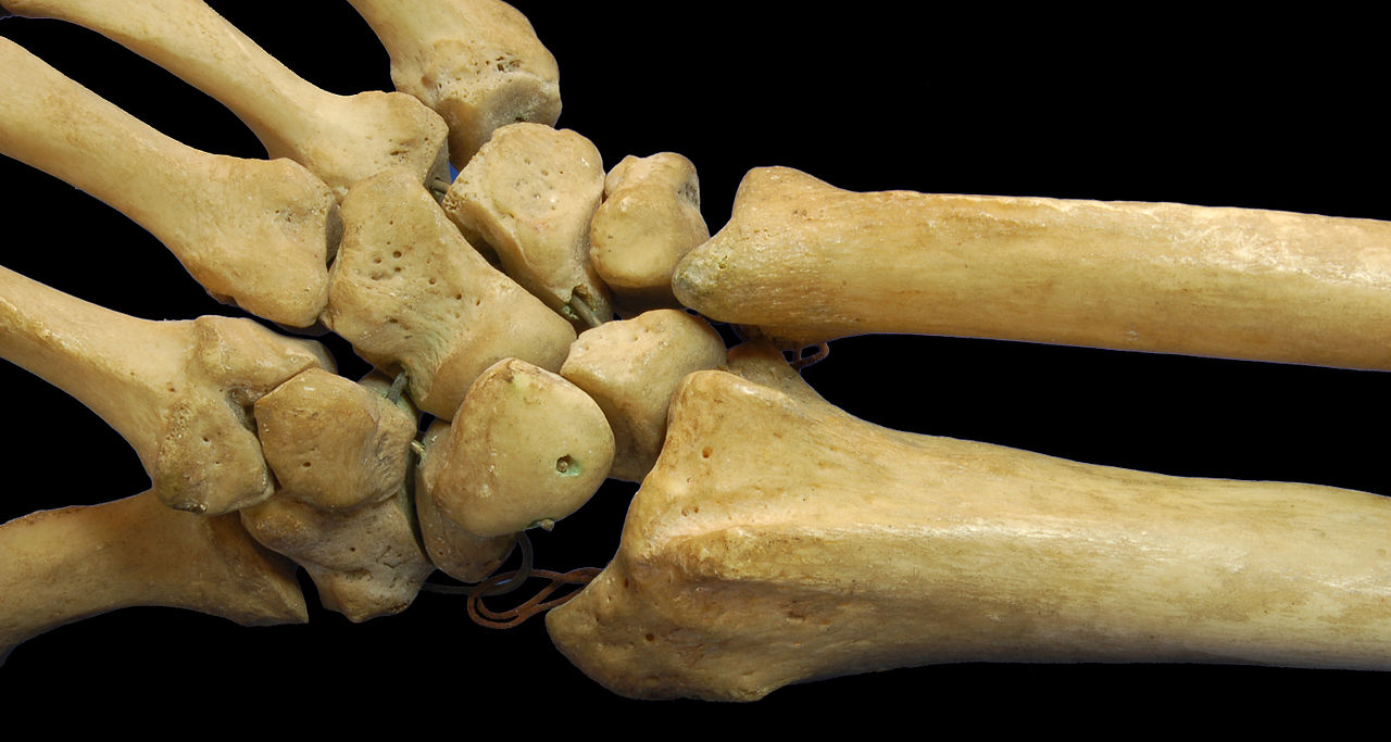
- Short Bones: cube-shaped bones that contain both compact and spongy bone, primarily spongy (Examples: carpals (wrist bones), tarsals (foot bones))
- Flat Bones: thin bones consisting of spongy bone sandwiched between compact bone (Examples: bones of the skull)
- Scapulae (shoulder blades)
- Sternum
- Rib bones
- Cranium (skull bones)
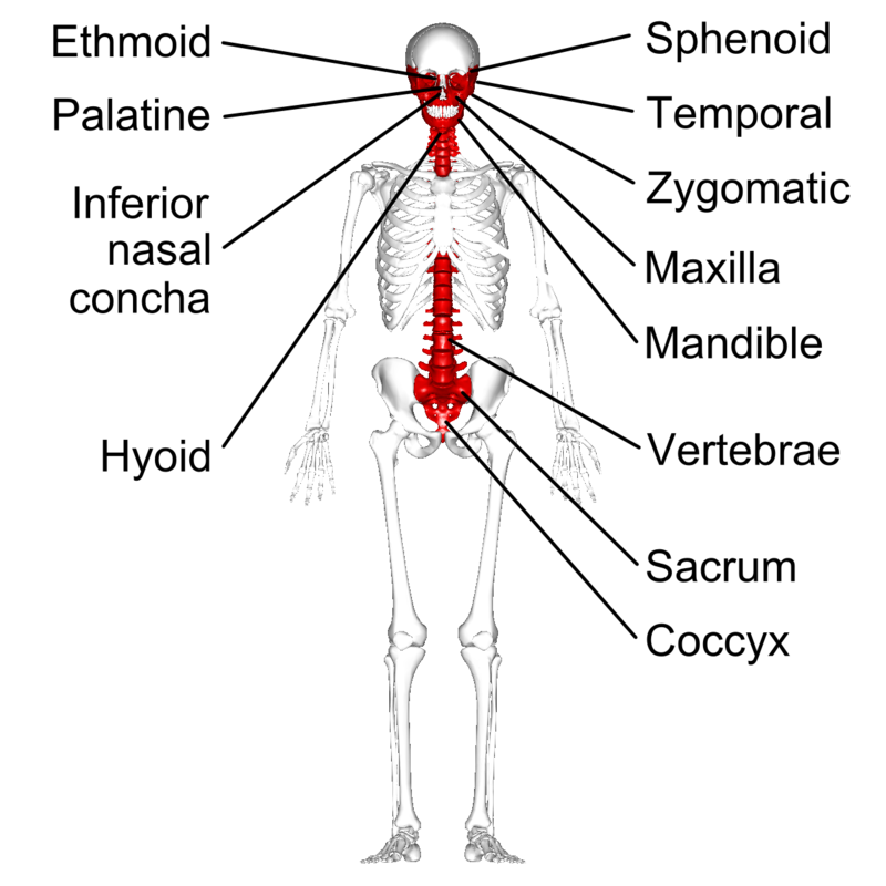
- Irregular Bones: irregular-shaped bones (Examples: the vertebrae of the spinal column and facial bones)
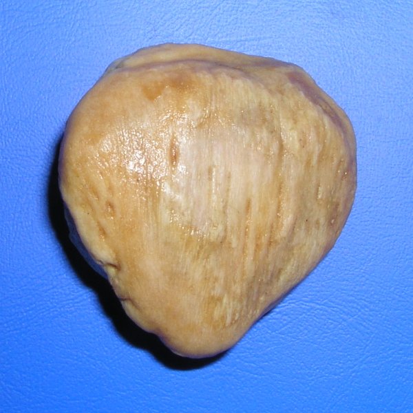
- Sesamoid Bone: round bone (Example: patalla/knee cap)
- Long Bones: mostly compact bone consisting of a shaft with heads at both ends (Examples: femur (thigh bone), humerus (arm bone), phalanges (fingers and toes))
Types of Bones:

https://upload.wikimedia.org/wikipedia/commons/thumb/7/77/Blausen_0229_ClassificationofBones.png/800px-Blausen_0229_ClassificationofBones.png
Flat Bones:
By OpenStax College - Anatomy & Physiology, Connexions Web site. http://cnx.org/content/col11496/1.6/, Jun 19, 2013., CC BY 3.0, https://commons.wikimedia.org/w/index.php?curid=30131423
https://upload.wikimedia.org/wikipedia/commons/thumb/d/db/RightHumanPosteriorDistalRadiusUlnaCarpals.jpg/1280px-RightHumanPosteriorDistalRadiusUlnaCarpals.jpg
https://upload.wikimedia.org/wikipedia/commons/1/18/Patella_ant.jpg
https://upload.wikimedia.org/wikipedia/commons/thumb/0/04/Irregular_bones_-_anterior_view_-_with_legend.png/800px-Irregular_bones_-_anterior_view_-_with_legend.png
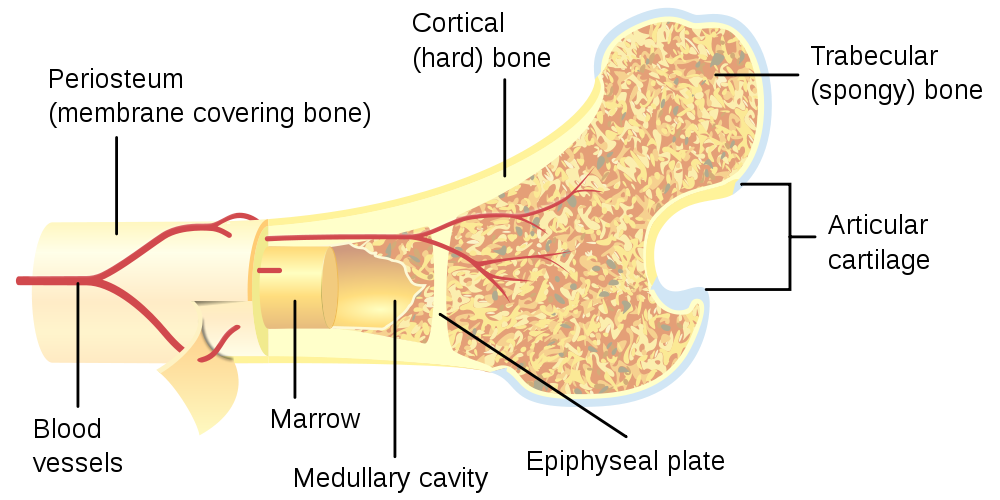
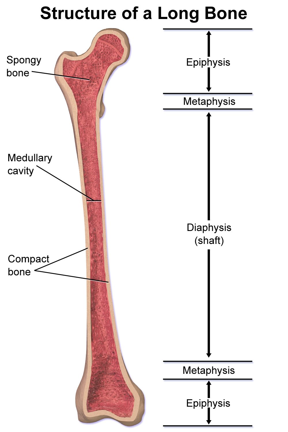
EXAMINATION OF A BONE: A LONG BONE
|
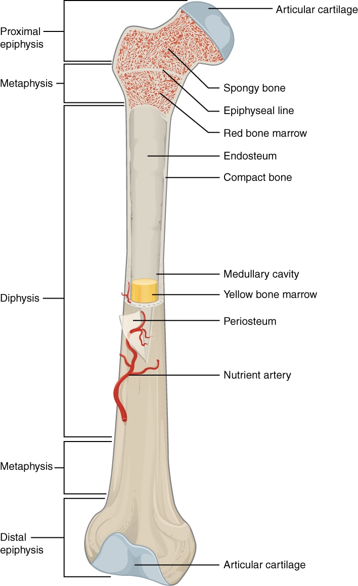
By OpenStax College - Anatomy & Physiology, Connexions Web site. http://cnx.org/content/col11496/1.6/, Jun 19, 2013., CC BY 3.0, https://commons.wikimedia.org/w/index.php?curid=30131409
|
By Pbroks13 - Own work, CC BY 3.0, https://commons.wikimedia.org/w/index.php?curid=5188772
https://upload.wikimedia.org/wikipedia/commons/f/fa/Structure_of_a_Long_Bone.png
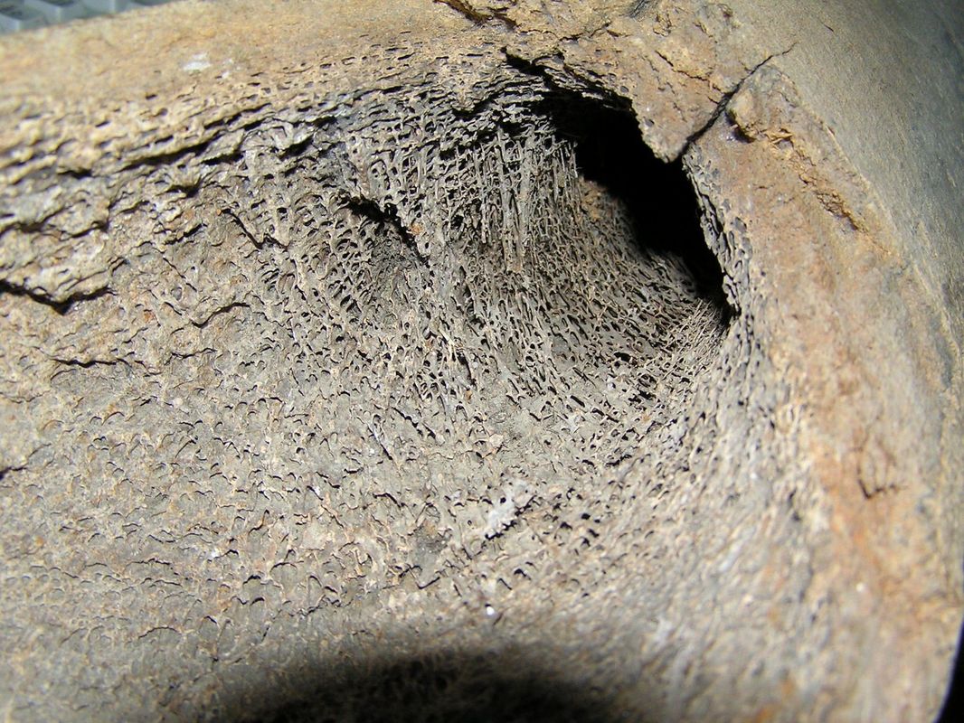
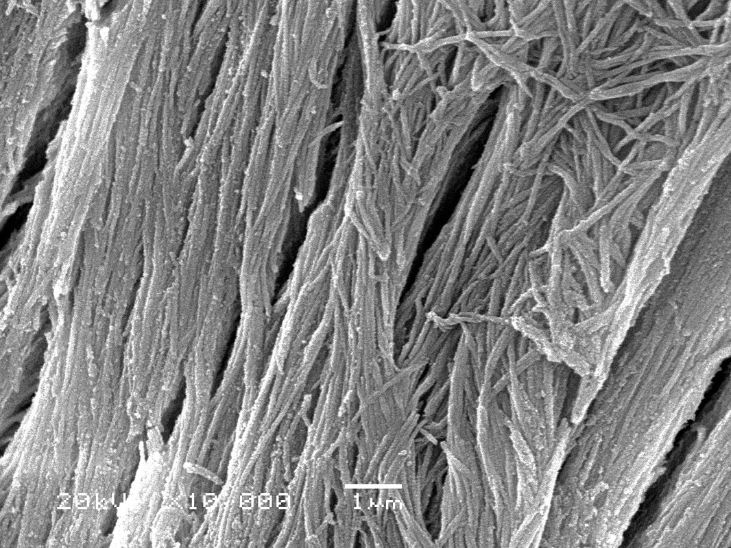
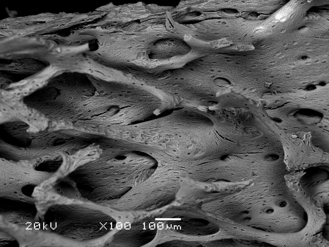
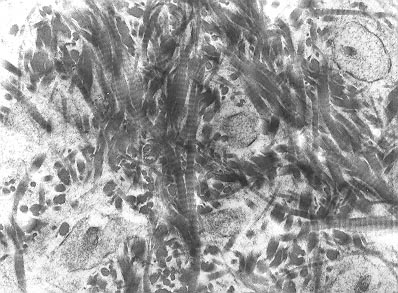
MICROSCOPIC EXAMINATION OF BONE:
|
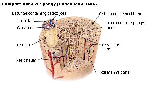
By SEER - U.S. National Cancer Institute's Surveillance, Epidemiology and End Results (SEER) Program (http://training.seer.cancer.gov/index.html)Exact adress, Public Domain, https://commons.wikimedia.org/w/index.php?curid=378948
|
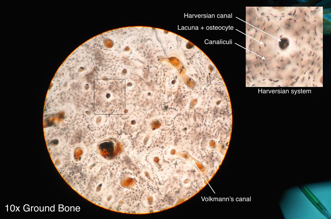
By Athikhun.suw - Own work, CC BY-SA 4.0, https://commons.wikimedia.org/w/index.php?curid=42368633
|
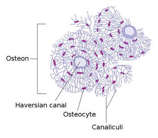
By Source digital bitmap graphics: BDBRecreated in vector format: Nyq - Original analog graphics: Gray’s Anatomy of the Human Body from the classic 1918 publication available online at Bartleby.com.Digital bitmap graphics: Transverse Section Of Bone.pngRecreated in vector format: Own work, CC BY-SA 4.0, https://commons.wikimedia.org/w/index.php?curid=50064939
|
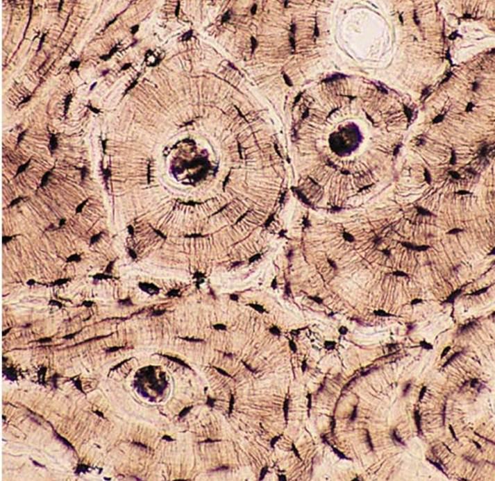
https://upload.wikimedia.org/wikipedia/commons/0/02/Bone_connective_tissue.jpg
By Daniel Ullrich Threedots - Own work, CC BY-SA 3.0, https://commons.wikimedia.org/w/index.php?curid=225845
|
By Sbertazzo - Own work, CC BY-SA 3.0, https://commons.wikimedia.org/w/index.php?curid=20904735
|
By Sbertazzo - Own work, CC BY-SA 3.0, https://commons.wikimedia.org/w/index.php?curid=20904767
|
Chemical Composition:
|
By Robert M. Hunt - Original work of Robert M. Hunt, Public Domain, https://commons.wikimedia.org/w/index.php?curid=13352900
|
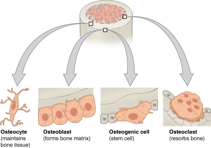
BONE CELLS, DEVELOPMENT, REMODELING:
- Osteocytes:
- Involved in reabsorption of bone tissue
- Mostly inactive osteoblasts trapped inside the matrix
- Occupy the lacunae (spaces)
- They have "processes" that reach out and interact with other bone cells
- Osteoblasts: these cells are the cells that make new bone
- Deposit a collagen matrix
- Release minerals
- Matrix and minerals combine to form new bone (creation and mineralization of new bone tissue)
- Mononucleate (single nucleus)
- Located on osteon surface
- Secrete a protein mixture (osteoid) mostly comprised of Type I collagen
- Manufacture hormones called prostaglandins to act on bone
- They secrete the enzyme alkaline phosphatase to mineralize bone and proteins
- Osteoclasts: These are the cells that breakdown bone
- Involved in reabsorption of bone tissue
- Bone is consistently remodeled by work of the osteoclasts and osteoblasts
- Multinucleate
- Located on resorption pits
- Form from monocyte stem cells
- Contain phagocytes similar to macrophages
- Mature and migrate to other bone surfaces
- It secretes enzymes such as tartrate resistant acid phosphatase
- Maintains calcium homeostasis (if not, bone spurs form)
By OpenStax College - Anatomy & Physiology, Connexions Web site. http://cnx.org/content/col11496/1.6/, Jun 19, 2013., CC BY 3.0, https://commons.wikimedia.org/w/index.php?curid=30131411
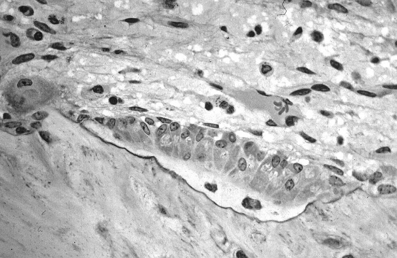
Osteoblasts and Osteocytes, By Robert M. Hunt - Own work, CC BY-SA 3.0, https://commons.wikimedia.org/w/index.php?curid=4230457
|
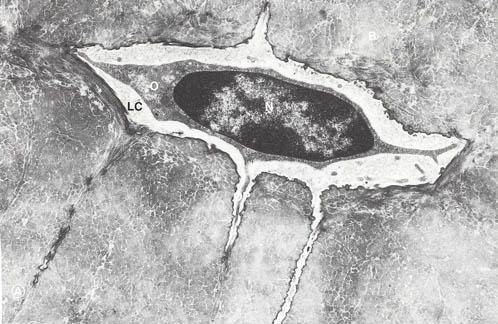
Osteocyte, By http://www.visualhistology.com/Visual_Histology_Atlas/VHA_Chpt6_Bone.html, Public Domain, https://commons.wikimedia.org/w/index.php?curid=3642719
|
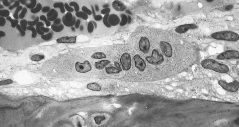
Osteoclasts, By Robert M. Hunt at English Wikipedia - Transferred from en.wikipedia to Commons by Kauczuk using CommonsHelper., Public Domain, https://commons.wikimedia.org/w/index.php?curid=7168671
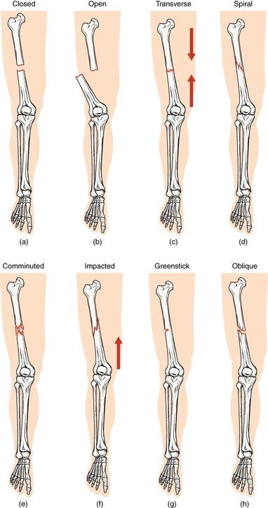
Types of Fractures:
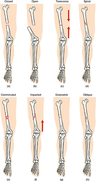
https://upload.wikimedia.org/wikipedia/commons/thumb/3/35/612_Types_of_Fractures.jpg/320px-612_Types_of_Fractures.jpg
Healing of Fractures:
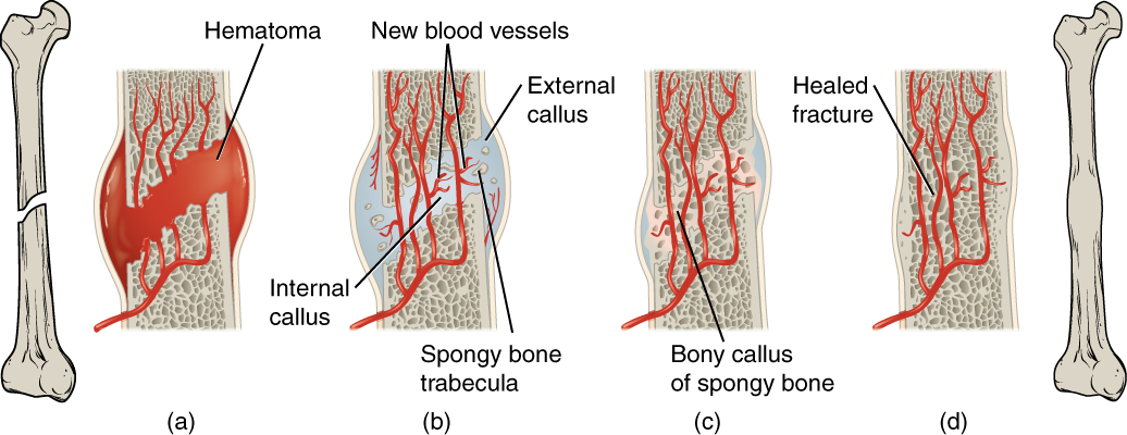
The Stages of Fracture Healing: https://upload.wikimedia.org/wikipedia/commons/1/12/613_Stages_of_Fracture_Repair.jpg
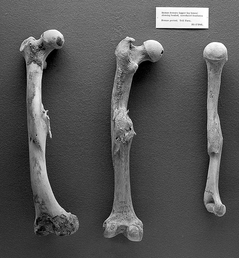
Healed bones from the Roman Period:
https://upload.wikimedia.org/wikipedia/commons/thumb/3/3a/Paleopathology%3B_Human_femurs_from_Roman_period%2C_Tell_Fara_Wellcome_L0008764.jpg/800px-Paleopathology%3B_Human_femurs_from_Roman_period%2C_Tell_Fara_Wellcome_L0008764.jpg
Soft Bone: Rickets
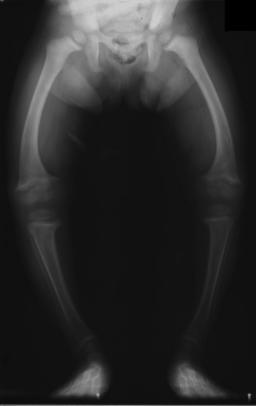
https://upload.wikimedia.org/wikipedia/commons/a/a9/XrayRicketsLegssmall.jpg
Osteoporosis (Brittle Bone):
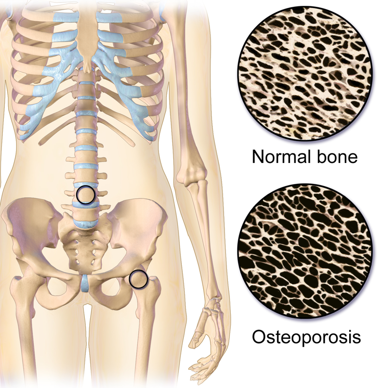
https://upload.wikimedia.org/wikipedia/commons/thumb/a/af/Osteoporosis_Locations.png/800px-Osteoporosis_Locations.png
|
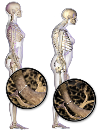
https://upload.wikimedia.org/wikipedia/commons/d/da/Blausen_0686_Osteoporosis_01.png
|
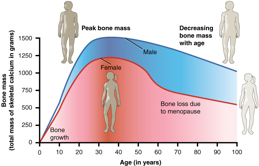
https://upload.wikimedia.org/wikipedia/commons/9/91/615_Age_and_Bone_Mass.jpg
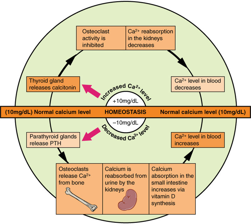
https://upload.wikimedia.org/wikipedia/commons/thumb/0/0b/625_Calcium_Homeostasis.jpg/800px-625_Calcium_Homeostasis.jpg
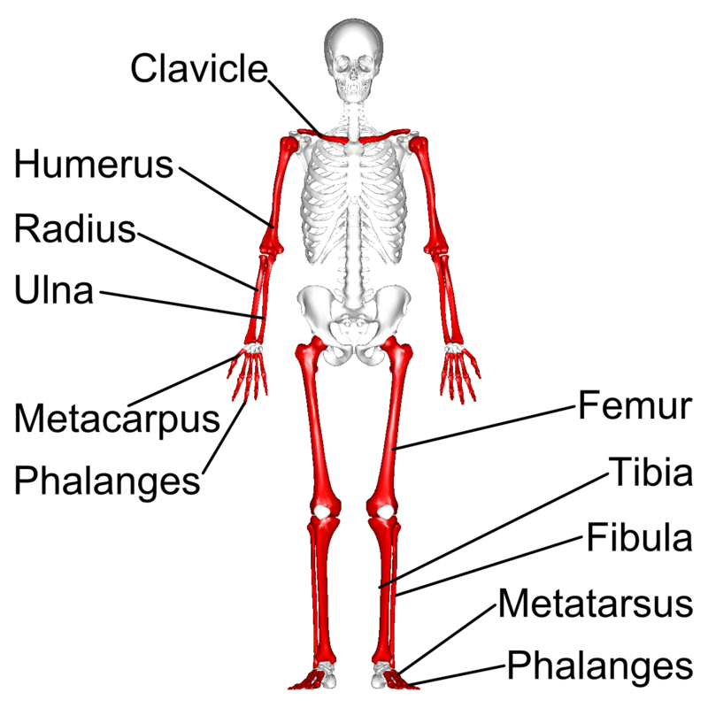
Images: Classification of Bone Types
By BruceBlaus - Own work, CC BY 3.0, https://commons.wikimedia.org/w/index.php?curid=29849179
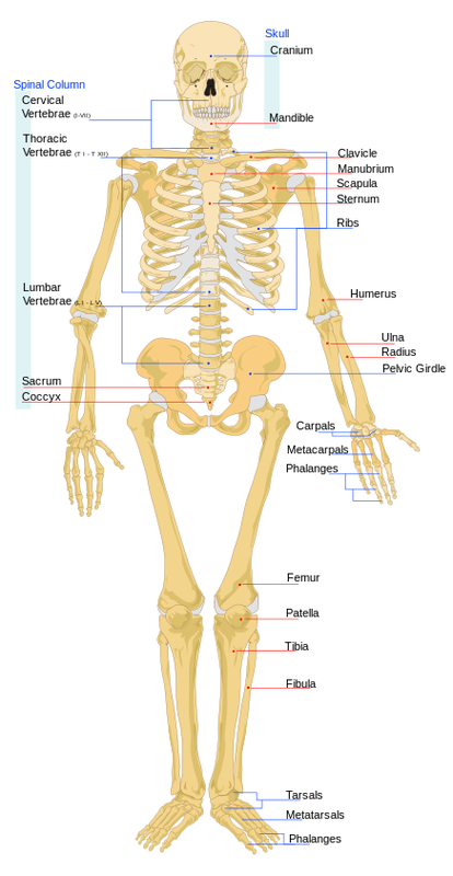
THE SKELETAL SYSTEM:
- The skeleton is made up of both cartilage and bone
- The embryo skeleton is made up mostly of hyaline cartilage, most of which later hardens by mineral salts and converts into bone or is replaced by harder bone tissue
- The skeletal tissue provides framework and support and protection for internal organs
- The bones function as levers for musculoskeletal movement
- Bone is a major storage center for minerals and mineral salts and even lipids
- Bone is a major site of blood cell production (occurs in red marrow cavities)
- Bones connect to other bones at joints (called articulations)
- There are two major sections to the skeleton: the appendicular and the axial skeletons
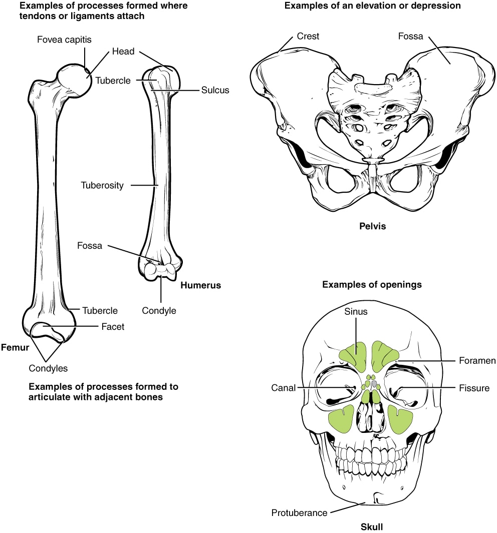
- Landmarks: Bony landmarks provide a map and are sometimes also referred to as bone markings
- Projections (processes)-sites of muscle or tendon attachment; some help anchor/form joints
- Depressions (cavities)-openings in bone that act as tunnels or indentations for nerves and blood supply
THE SKELETON: APPENDICULAR AND AXIAL

APPENDICULAR SKELETON;By Anatomography is provided by DBCLS - en:Anatomography (setting page of this image at Anatomography), CC BY-SA 2.1 jp, https://commons.wikimedia.org/w/index.php?curid=38025137
|

AXIAL SKELETON;By Anatomography is provided by DBCLS - en:Anatomography (setting page of this image at Anatomography), CC BY-SA 2.1 jp, https://commons.wikimedia.org/w/index.php?curid=38024088
|
The Appendicular Skeleton:
- This portion of the skeleton consists of the appendages (arm and leg bones, hand and foot bones), pectoral (shoulder bones, scapulae, collar bone) and pelvic girdle (pelvic and hip bones)
- There are 126 bones in this portion of the skeleton
- Appendages are also called limbs
- They are connected by freely movable joints
Objectives:
Materials:
- Identify the principal bone markings of the pectoral (shoulder) girdle and the upper limb
- Describe the articulation of the pectoral girdle with the humerus
- Describe the articulation of the humerus with the forearm bones
- Identify the bone markings of the pelvic girdle and the lower limbs
- Describe the articulation of the bones of the pelvic girdle with the femur
- Describe the articulation of the femur with the leg bones
Materials:
- Lab Manual
- Skeleton and Carolina Biological Distance Learning Lab Kit (Musculoskeletal System)
- Appendere = to add something
- Has larger bones than the axial skeleton
- Bears more weight than the axial skeleton
- Composed of 126 bones
- Make up the upper and lower limbs or extremities and bones or girdles that attach the limbs to the axial skeleton
- 4 main areas:
- Pectoral girdles
- Pectus = breast
- Girdle = any curved structure
- Upper limbs
- Pelvic girdle
- Pelvis = basin
- Lower limbs
- Pectoral girdles
- Bone markings: include sites of attachment of muscles and articulations with other bones to form a joint (see Table 10.1):
- Crest
- Condyle
- Epicondyle
- Foramen
- Fossa
- Head
- Notch
- Trochanter
- Spine
- Tuberosity
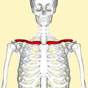
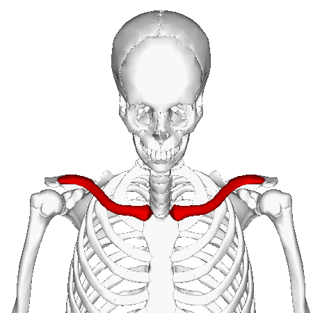
- Two pectoral girdles, each attaching to an upper limb to the axial skeleton
- Pectoral girdle (shoulder girdle):
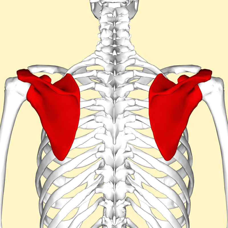
- Scapula (shoulder blade)
- Spine - sharp ridge located on posterior side
- Acromion - flattened process at the lateral end of the spine that articulates with the clavicle; also called the acromial process
- Glenoid cavity - depression (fossa) inferior to acromion that articulates with the head of the humerus
- Glene = joint socket
- Coracoid process - superior and medial to the glenoid cavity; site for muscle attachment
- Coracoid = crow's beak
- Clavicle (key) - collar bone
- Scapula (shoulder blade)

By BruceBlaus. When using this image in external sources it can be cited as:Blausen.com staff (2014). "Medical gallery of Blausen Medical 2014". WikiJournal of Medicine 1 (2). DOI:10.15347/wjm/2014.010. ISSN 2002-4436. - Own work, CC BY-SA 3.0, https://commons.wikimedia.org/w/index.php?curid=27700914
The Pectoral Girdle:
Functions:
Upper Limb:
The Arm Bones:
|
By Anatomography - en:Anatomography (setting page of this image), CC BY-SA 2.1 jp, https://commons.wikimedia.org/w/index.php?curid=23923969
By Anatomography - en:Anatomography (setting page of this image), CC BY-SA 2.1 jp, https://commons.wikimedia.org/w/index.php?curid=23923926
By Anatomography - en:Anatomography (setting page of this image), CC BY-SA 2.1 jp, https://commons.wikimedia.org/w/index.php?curid=23378227
By Anatomography - en:Anatomography (setting page of this image), CC BY-SA 2.1 jp, https://commons.wikimedia.org/w/index.php?curid=23389521
By Anatomography - en:Anatomography (setting page of this image), CC BY-SA 2.1 jp, https://commons.wikimedia.org/w/index.php?curid=23590243
By Anatomography - en:Anatomography (setting page of this image), CC BY-SA 2.1 jp, https://commons.wikimedia.org/w/index.php?curid=24764500
By BruceBlaus. When using this image in external sources it can be cited as:Blausen.com staff (2014). "Medical gallery of Blausen Medical 2014". WikiJournal of Medicine 1 (2). DOI:10.15347/wjm/2014.010. ISSN 2002-4436. - Own work, CC BY 3.0, https://commons.wikimedia.org/w/index.php?curid=29849185
|
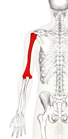
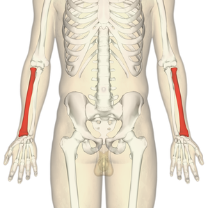
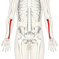
Bones and Selected Bone Markings of the Arm and Forearm:
Humerus (Upper Arm Bone)
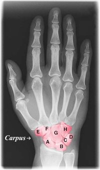
Carpals (Wrists)
Humerus (Upper Arm Bone)
- Head
- Deltoid tuberosity
- Trochlea
- Capitulum
- Coronoid fossa
- Olecranon fossa
- Olecranon
- Coronoid process
- Trochlear notch
- Styloid process
- Head
- Styloid process
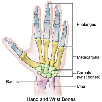
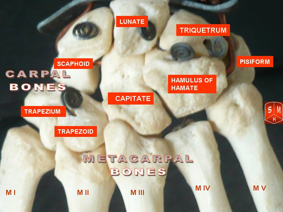
Carpals (Wrists)
- Carpal bones: 8 short bones of the wrist
- Metacarpal bones: 5 bones that make up the palm of the hand, numbered I to V
- Phalanges: (singular: phalanx): Bones that make up the fingers numbered I to V: Proximal, Middle, Distal
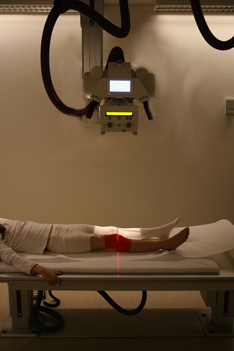
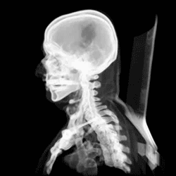
Radiography is used to take X-ray images of a bone.
By Anatomist90 - Own work, CC BY-SA 3.0, https://commons.wikimedia.org/w/index.php?curid=17406951
|
By Original photo is by Dr. Jochen Lengerke at de.wikipedia. Painted color by User:Was a bee. - File:Xray hand.jpg, Public Domain, https://commons.wikimedia.org/w/index.php?curid=30080248
|
X-Rays:
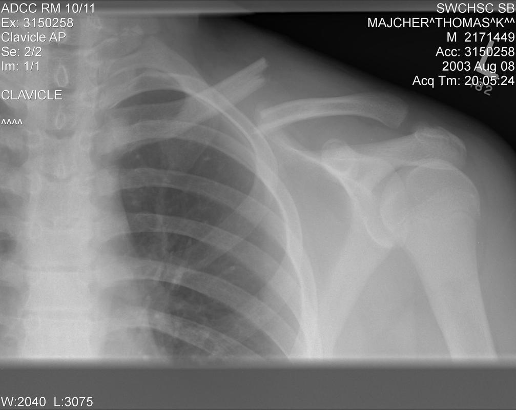
Broken clavicle (collar bone) is a common sports injury, particularly in football;By Majorkev (talk) (Uploads) - Own work, CC BY 3.0, https://en.wikipedia.org/w/index.php?curid=39495972
|
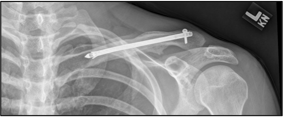
Broken clavicle bone surgical repair;By Bentplate - Own work, CC BY-SA 3.0, https://commons.wikimedia.org/w/index.php?curid=16225724
|
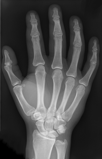
By Hellerhoff - Own work, CC BY-SA 3.0, https://commons.wikimedia.org/w/index.php?curid=12143891; hand bones
|
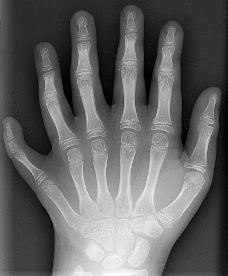
Polydactyly: An extra finger (3 phalanges, a metacarpal);By en:User:Drgnu23, subsequently altered by en:user:Grendelkhan, en:user: Raul654, and en:user:Solipsist. - Own work, CC BY-SA 3.0, https://commons.wikimedia.org/w/index.php?curid=120504
|
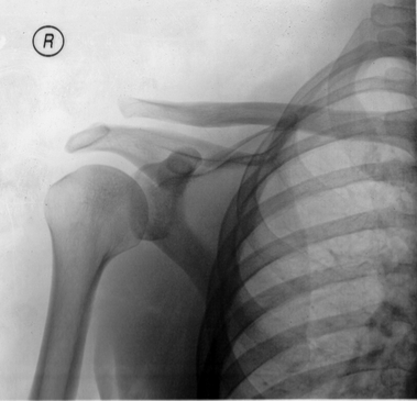
An AC joint separation, By Majorkev (talk) (Uploads) - Own work, CC BY 3.0, https://en.wikipedia.org/w/index.php?curid=39495972
|
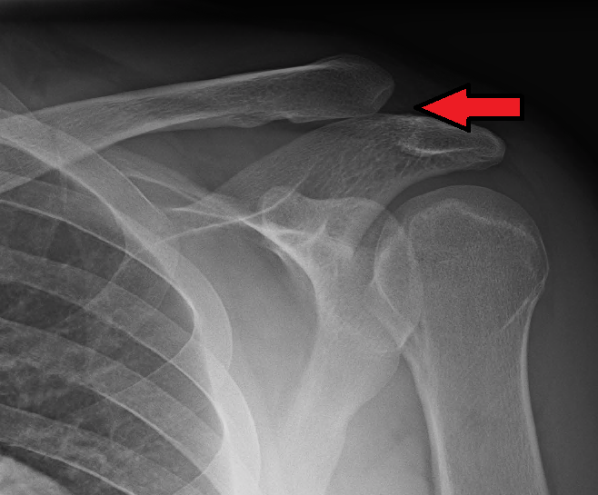
Grade 3 AC joint separation
|
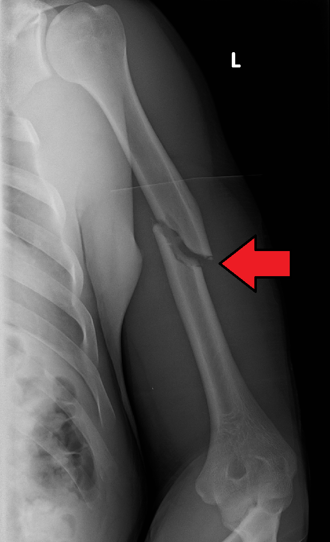
Humerus fracture;By James Heilman, MD - Own work, CC BY-SA 4.0, https://commons.wikimedia.org/w/index.php?curid=49147236
|
Types of fractures:By OpenStax College - Anatomy & Physiology, Connexions Web site. http://cnx.org/content/col11496/1.6/, Sep 7, 2015., CC BY 4.0, https://commons.wikimedia.org/w/index.php?curid=30127535
|
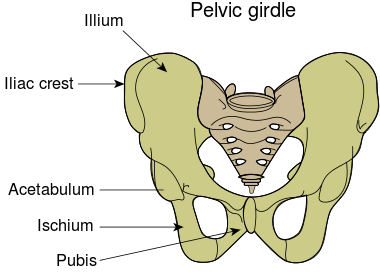
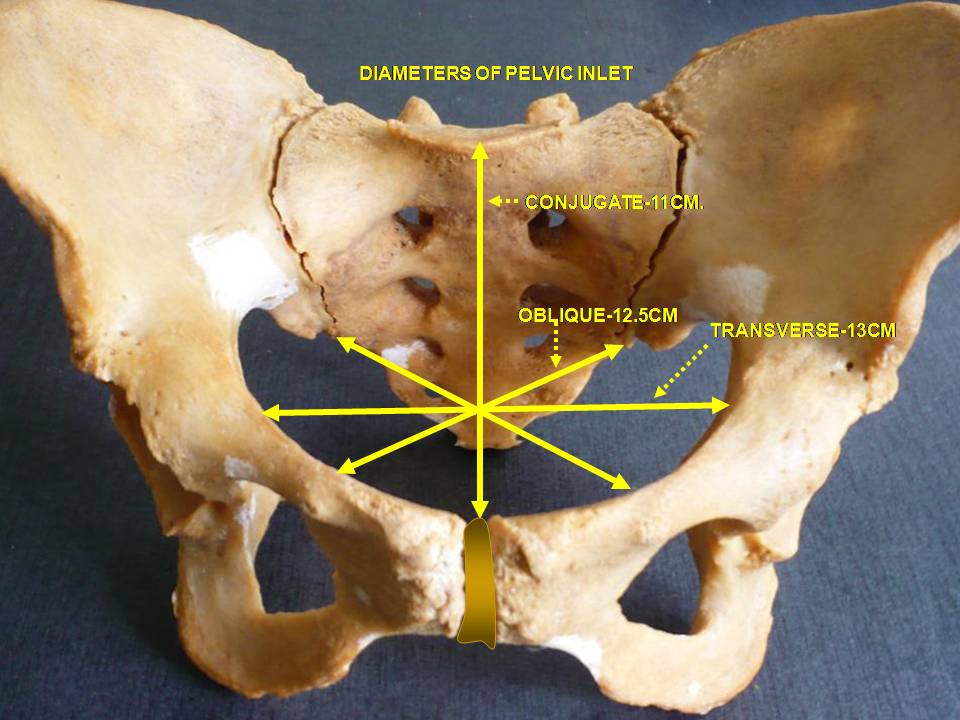
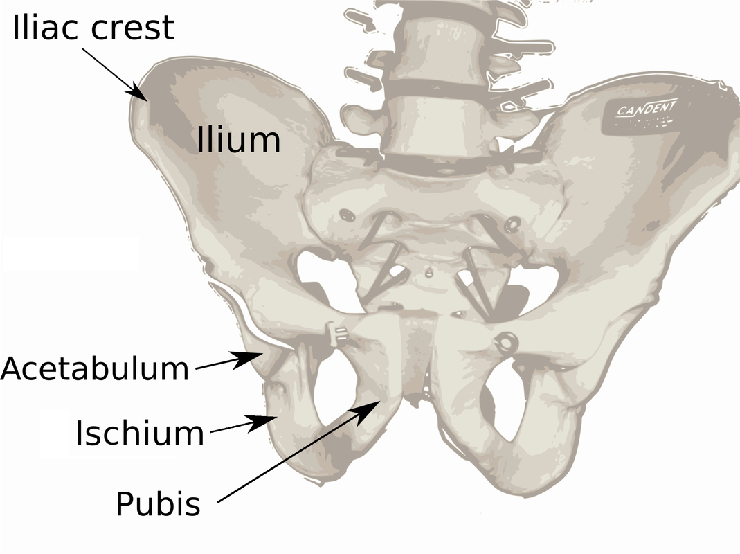
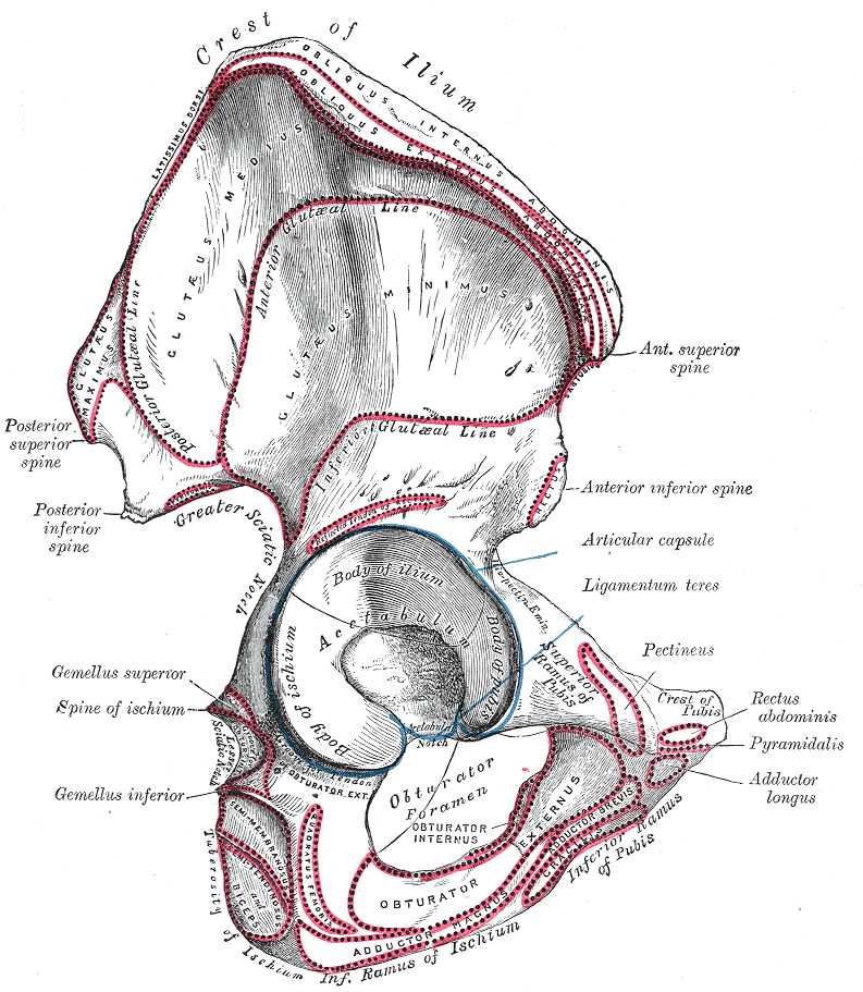
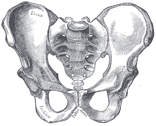
The Pelvic Girdle:
Pelvic Girdle:
- Composed of 2 hip or coxal bones (ox coxae) that attach the lower limb to the axial skeleton
- Os coxa (singular) - hip bone: formed by the fusion of 3 bones:
- Ilium - largest and most superior bone
- Iliac crest - superior border of ilium; when placing hands on "hips", you're on this area
- Anterior superior iliac spine - protrusion on anterior end of iliac crest
- Greater sciatic notch - large notch on posterior side
- Ischium - inferior, posterior portion of the os coxa; you sit on this bone
- Pubis - anterior, inferior portion of os coxa
- Pubis symphysis - joint where the 2 pubic bones join anteriorly
- Bone Markings:
- Acetabulum - cup-shaped indentation for the head of the femur; forms the hip joint (coxal joint) - a ball-and-socket joint connected by a strong ligament
- Obturator foramen - largest foramen in the skeleton
- Sacroiliac joint - os coxae jointed posteriorly with the sacrum here, forming the bony pelvis that connects the lower extremity with the axial skeleton
- Ilium - largest and most superior bone
By Original: U.S. National Cancer Institute; Vectorization: Fred the Oyster; German translation kopiersperre/Rothwild - Own work based on: Illu pelvic girdle.jpg, CC BY-SA 4.0, https://commons.wikimedia.org/w/index.php?curid=35388599

By Fred the Oyster, CC BY-SA 4.0, https://commons.wikimedia.org/w/index.php?curid=35384964;
1. Sacrum
2. Ilium
3. Ischium
4. Pubic Bone
5. Pubis Symphysis Joint
6. Acetabulum
7. Foramen Obturatum
8. Coccyx (Tailbone)
By Anatomist90 - Own work, CC BY-SA 3.0, https://commons.wikimedia.org/w/index.php?curid=23305501
By Je at uwo at English Wikipedia - Transferred from en.wikipedia to Commons., Public Domain, https://commons.wikimedia.org/w/index.php?curid=2289285
|
|
Some Types of Low Back Pain/Pelvic Pain:
|
By Henry Vandyke Carter - Henry Gray (1918) Anatomy of the Human Body (See "Book" section below)Bartleby.com: Gray's Anatomy, Plate 235, Public Domain, https://commons.wikimedia.org/w/index.php?curid=792148
|
The Male Versus Female Pelvic Girdle:
The Male Pelvis:By Henry Vandyke Carter - Henry Gray (1918) Anatomy of the Human Body (See "Book" section below)Bartleby.com: Gray's Anatomy, Plate 241, Public Domain, https://commons.wikimedia.org/w/index.php?curid=323668
|
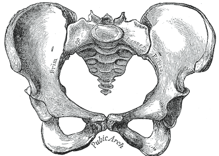
The Female Pelvis:By Henry Vandyke Carter - Henry Gray (1918) Anatomy of the Human Body (See "Book" section below)Bartleby.com: Gray's Anatomy, Plate 242, Public Domain, https://commons.wikimedia.org/w/index.php?curid=323669
|
|
|
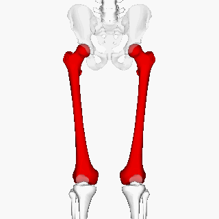
The Thigh and Leg Bones:
Lower Limbs: 30 bones (4 thigh & leg bones; 26 foot bones)
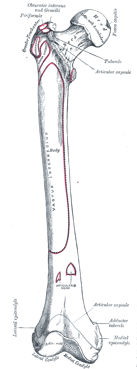
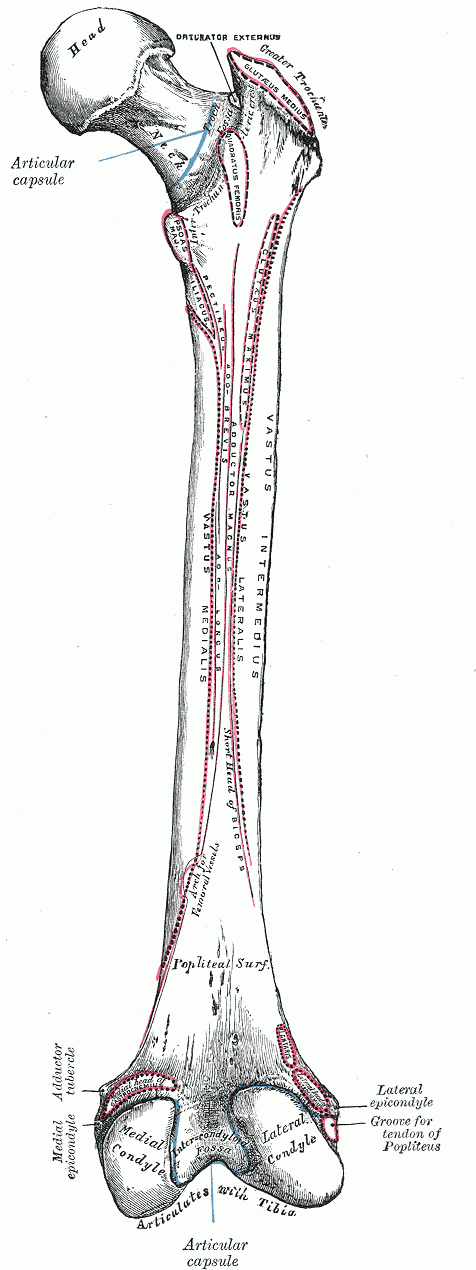
- Femur - thigh bone; largest and strongest bone in the human skeleton
- Head - large, rounded, knob-like proximal end
- Neck - narrower, constriction distal to head
- Greater trochanter - large and roughened superior projection lateral to neck
- Medial condyle - rounded, medial process on posterior side of distal end
- Lateral condyle - rounded, lateral process on posterior side of lateral end
- Patella - kneecap; small, sesamoid bone
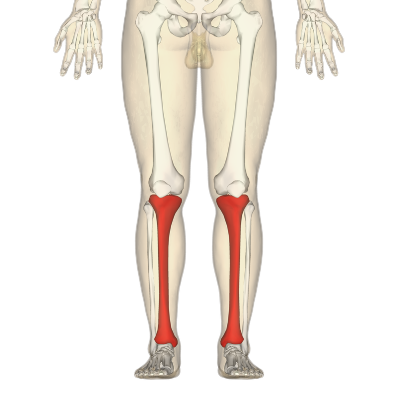
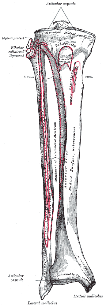
- Tibia - leg bone; weight-bearing; medially located; forms a joint laterally with the fibula
- Medial condyle - flattened, expanded medial projections on proximal end
- Lateral condyle - similar to medial condyle on lateral side
- Tibial tuberosity - large, roughened projection on anterior surface, inferior to condyles
- Medial malleolus - medial process on distal end, forms medial bump of ankle
- Fibula - leg bone; slender lateral leg bone that is important for muscle attachment, but not for bearing weight
- Head - proximal; articulates with the tibia but not the femur
- Lateral malleolus - distal and articulates with the talus laterally; forms the bump of the ankle
- Tarsals - ankle bones; composed of 7 bones, 2 of them being larger than the rest
- Calcaneous - heel bone
- Calcaneum = heel
- Talus - ankle bone that articulates with the tibia
- Talus = ankle
- Calcaneous - heel bone
- Metatarsals - Anterior portion of the instep, composed of 5 bones that coincide with the metacarpals in the hand, numbered I to V from the great toe to the little toe
- Meta = after or next
- Phalanges - toes or digits; similar to the phalanges of the hand, numbered I to V from the great toe to the little toe
- Great toe - made up to 2 phalanges: proximal and distal
- Digits II - V have 3 bones each: proximal, middle, distal phalange
The Thigh Bone (Femur):
|
By Anatomography - en:Anatomography (setting page of this image), CC BY-SA 2.1 jp, https://commons.wikimedia.org/w/index.php?curid=23812919
|
|
|
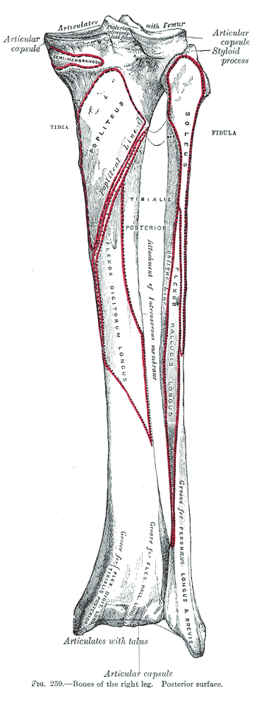
The Leg Bones: Tibia and Fibula
The Tibia: Shin Bone
|
By Anatomography - en:Anatomography (setting page of this image), CC BY-SA 2.1 jp, https://commons.wikimedia.org/w/index.php?curid=24693053
|
By Henry Vandyke Carter - Henry Gray (1918) Anatomy of the Human Body (See "Book" section below)Bartleby.com: Gray's Anatomy, Plate 258, Public Domain, https://commons.wikimedia.org/w/index.php?curid=792113
|
By Henry Vandyke Carter - Henry Gray (1918) Anatomy of the Human Body (See "Book" section below)Bartleby.com: Gray's Anatomy, Plate 259, Public Domain, https://commons.wikimedia.org/w/index.php?curid=792109
|
The Foot Bones:
|
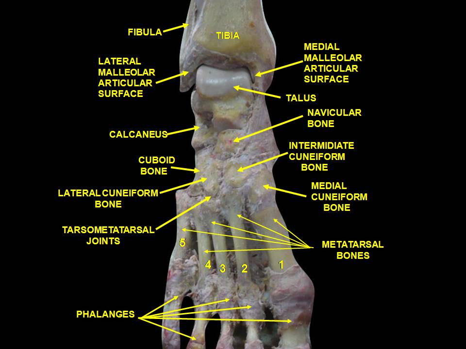
By Anatomist90 - Own work, CC BY-SA 3.0, https://commons.wikimedia.org/w/index.php?curid=25508267
|
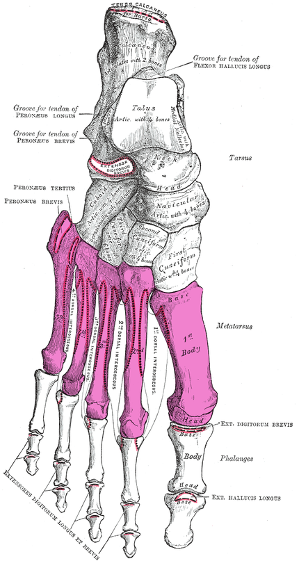
By Henry Vandyke Carter - Henry Gray (1918) Anatomy of the Human Body (See "Book" section below)Bartleby.com: Gray's Anatomy, Plate 268, Public Domain, https://commons.wikimedia.org/w/index.php?curid=28558193
|
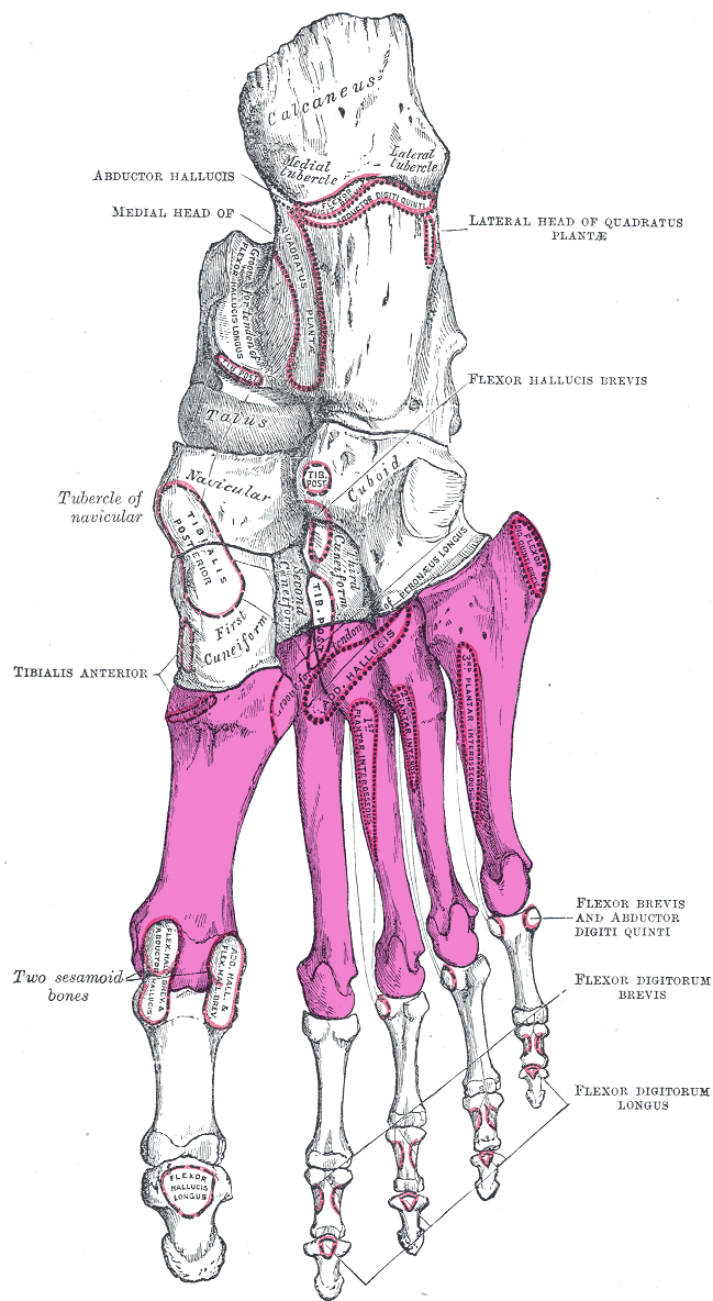
By Henry Vandyke Carter - Henry Gray (1918) Anatomy of the Human Body (See "Book" section below)Bartleby.com: Gray's Anatomy, Plate 269, Public Domain, https://commons.wikimedia.org/w/index.php?curid=28558459
|
The Axial Skeleton:

By BruceBlaus. When using this image in external sources it can be cited as:Blausen.com staff (2014). "Medical gallery of Blausen Medical 2014". WikiJournal of Medicine 1 (2). DOI:10.15347/wjm/2014.010. ISSN 2002-4436. - Own work, CC BY 3.0, https://commons.wikimedia.org/w/index.php?curid=27796925
- Consists of the skull, the vertebral column and the thorax
BONY LANDMARKS OR BONE MARKINGS:
|
Projections:
|
|
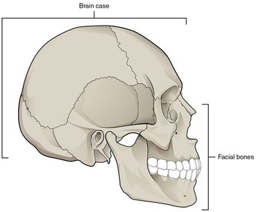
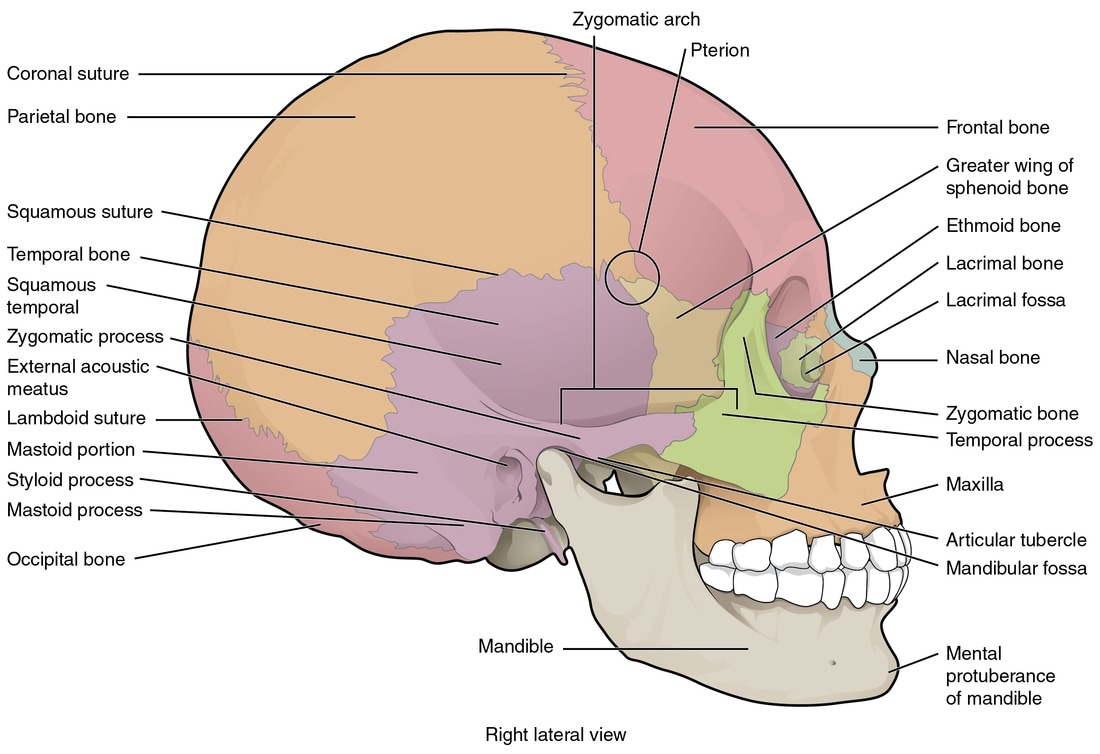
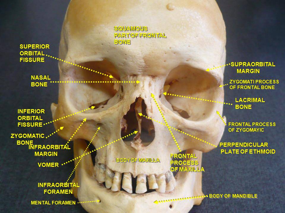
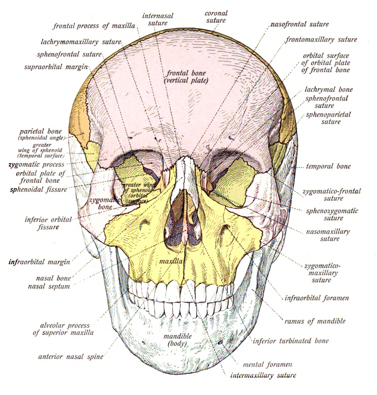
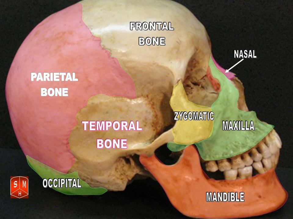
THE SKULL:
|
By Arielinson - Own work, CC BY-SA 4.0, https://commons.wikimedia.org/w/index.php?curid=50903553
|
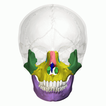
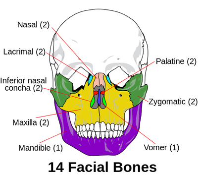
Craniofacial Bones:
By OpenStax College - Anatomy & Physiology, Connexions Web site. http://cnx.org/content/col11496/1.6/, Jun 19, 2013., CC BY 3.0, https://commons.wikimedia.org/w/index.php?curid=30131432
|
By OpenStax College - Anatomy & Physiology, Connexions Web site. http://cnx.org/content/col11496/1.6/, Jun 19, 2013., CC BY 3.0, https://commons.wikimedia.org/w/index.php?curid=30131435
|
By Polygon data is from BodyParts3D - Polygon data is from BodyParts3D, CC BY-SA 2.1 jp, https://commons.wikimedia.org/w/index.php?curid=37620562
|
By Own work - File:Es-Human skull front simplified (bones).svg, Public Domain, https://commons.wikimedia.org/w/index.php?curid=37631440
|
By Anatomist90 - Own work, CC BY-SA 3.0, https://commons.wikimedia.org/w/index.php?curid=30039012
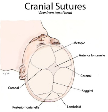
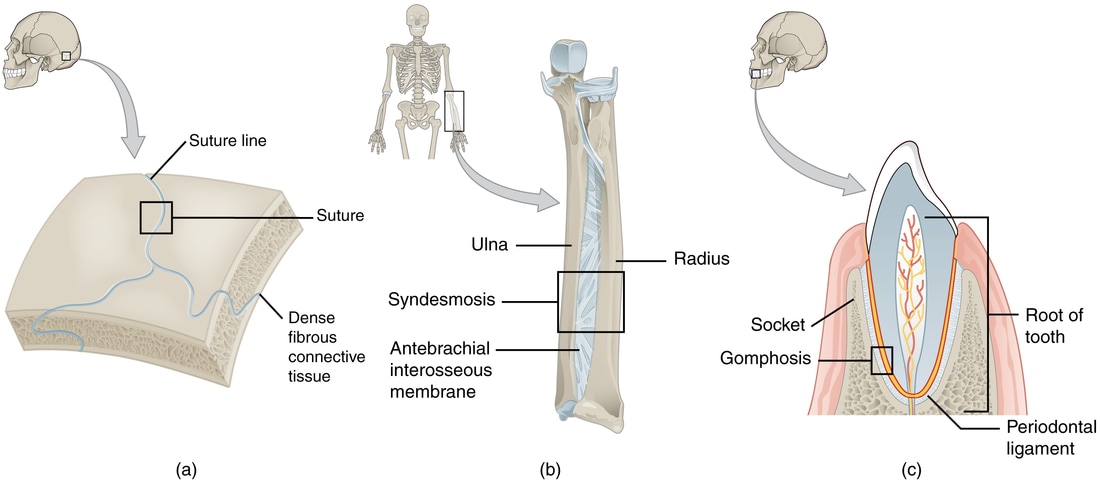
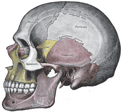
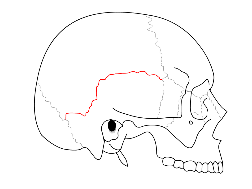
Fibrous Joints: Sutures
By OpenStax College - Anatomy & Physiology, Connexions Web site. http://cnx.org/content/col11496/1.6/, Jun 19, 2013., CC BY 3.0, https://commons.wikimedia.org/w/index.php?curid=30131529
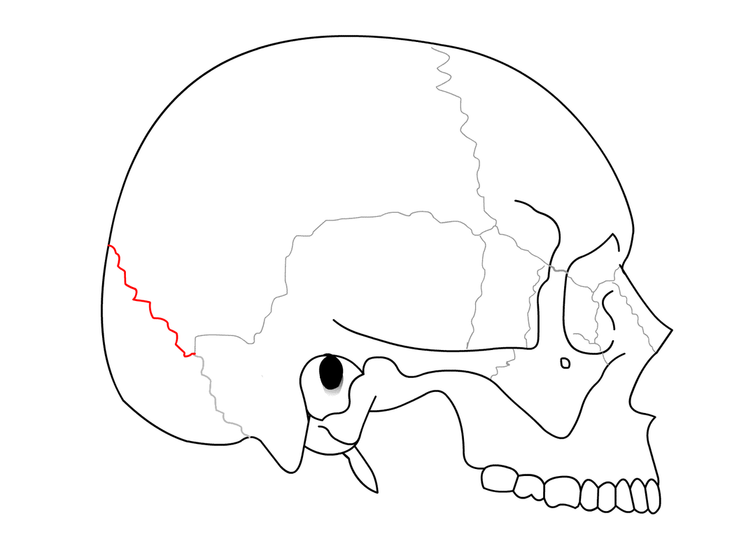
The Cranial Sutures:
By Henry Vandyke Carter - Henry Gray (1918) Anatomy of the Human Body (See "Book" section below)Bartleby.com: Gray's Anatomy, Plate 188, Public Domain, https://commons.wikimedia.org/w/index.php?curid=556832
|

By Xxjamesxx - Own work, CC BY-SA 3.0, https://commons.wikimedia.org/w/index.php?curid=12055702
|
Lambdoid Suture;By I, RosarioVanTulpe, CC BY-SA 3.0, https://commons.wikimedia.org/w/index.php?curid=24272529
|
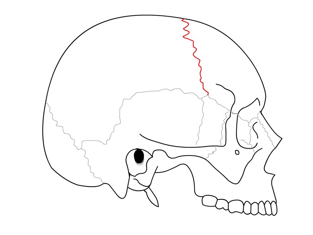
Coronal Suture;By I, RosarioVanTulpe, CC BY-SA 3.0, https://commons.wikimedia.org/w/index.php?curid=2421006
|
Squamous Suture;By I, RosarioVanTulpe, CC BY-SA 3.0, https://commons.wikimedia.org/w/index.php?curid=2421012
|
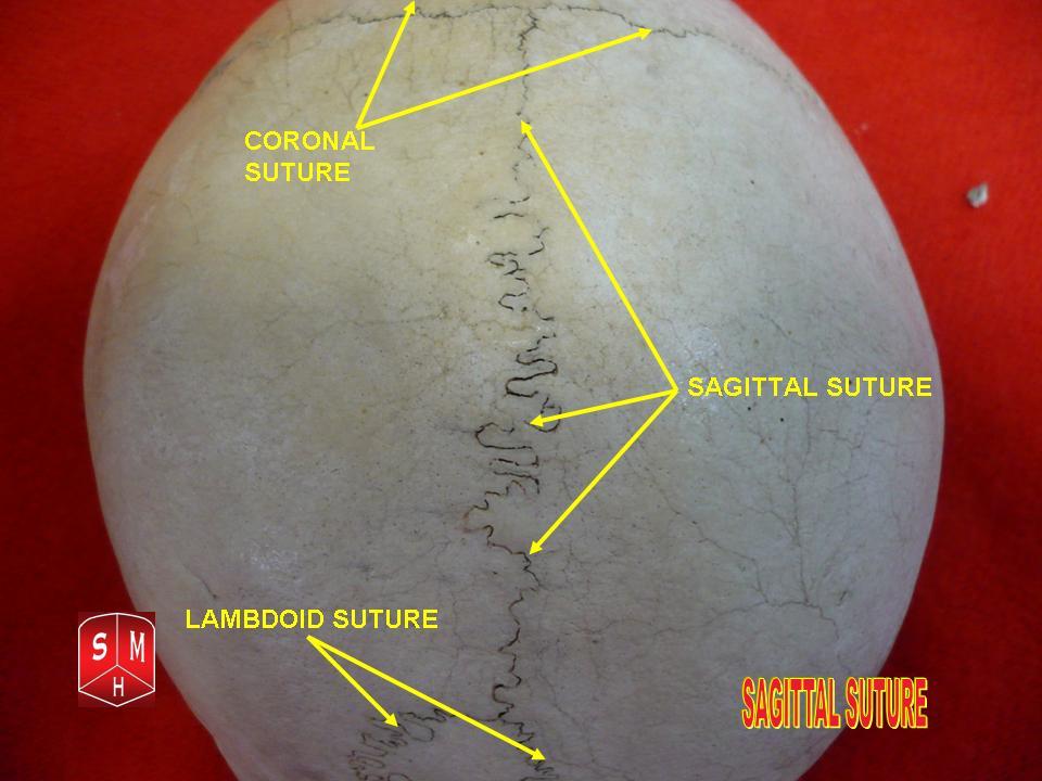
Sagittal Suture; By Anatomist90 - Own work, CC BY-SA 3.0, https://commons.wikimedia.org/w/index.php?curid=17553144
|
By Dr. Johannes Sobotta - Sobotta Atlas and Text-book of Human Anatomy 1909, Public Domain, https://commons.wikimedia.org/w/index.php?curid=29760948
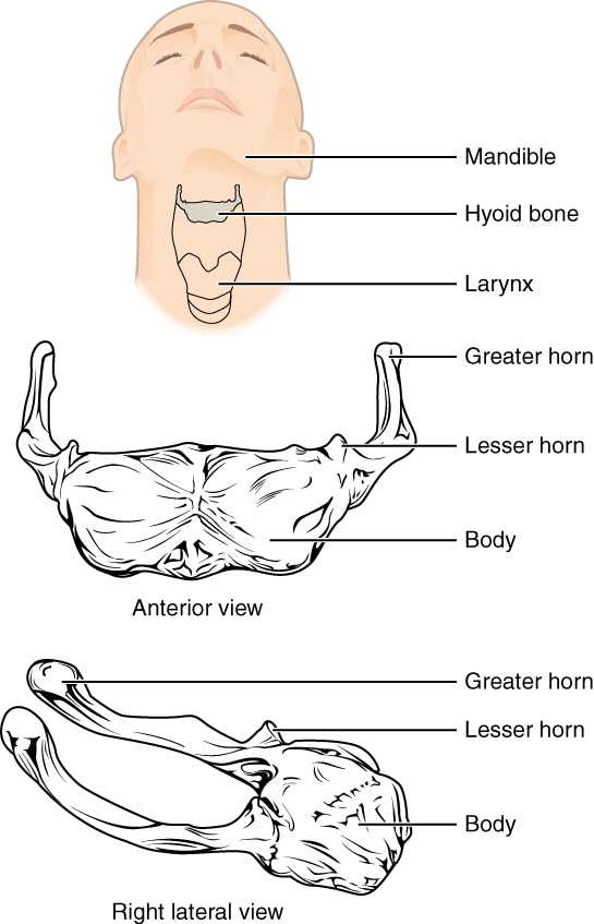
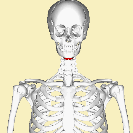
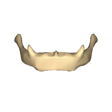
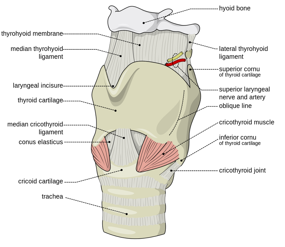
The Hyoid Bone:
By OpenStax College - Anatomy & Physiology, Connexions Web site. http://cnx.org/content/col11496/1.6/, Jun 19, 2013., CC BY 3.0, https://commons.wikimedia.org/w/index.php?curid=30131442
|
|
By Anatomography - en:Anatomography (setting page of this image), CC BY-SA 2.1 jp, https://commons.wikimedia.org/w/index.php?curid=24001578
|
By Anatomography - en:Anatomography (setting page of this image), CC BY-SA 2.1 jp, https://commons.wikimedia.org/w/index.php?curid=24001337
|
By Olek Remesz (wiki-pl: Orem, commons: Orem) - Own work, modified SVG version of PD picture from Gray's Anatomy., CC BY-SA 2.5-2.0-1.0, https://commons.wikimedia.org/w/index.php?curid=3492701
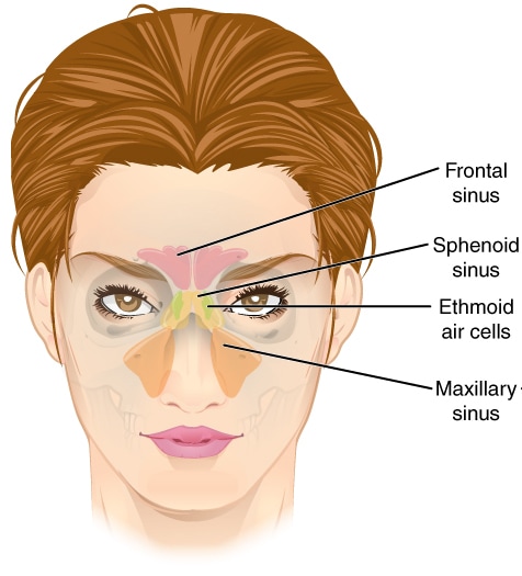
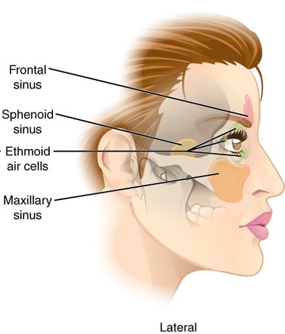
The Paranasal Sinuses:
- The sinuses are mucosal-lined air cavities:
- Frontal Sinus
- Sphenoid Sinus
- Ethmoid Sinus
- Maxillary Sinus
- Their purpose is to make the skull lighter
- They also serve to act as resonance chambers for speech and language
- The largest sinus: The maxillary sinus
By OpenStax College - Anatomy & Physiology, Connexions Web site. http://cnx.org/content/col11496/1.6/, Jun 19, 2013., CC BY-SA 3.0, https://commons.wikimedia.org/w/index.php?curid=30850944
|
By CFCF - Own work, CC BY-SA 3.0, https://commons.wikimedia.org/w/index.php?curid=30850945
|
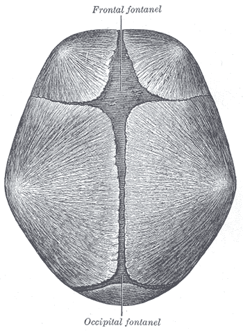
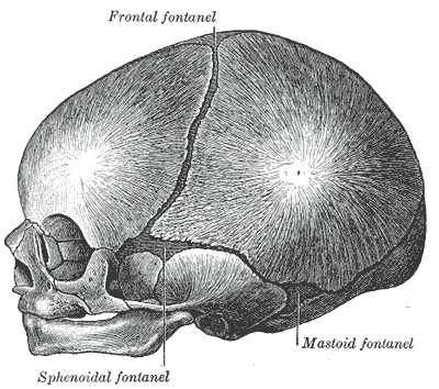
The Fetal Skull:
By CFCF - Own work, CC BY-SA 3.0, https://commons.wikimedia.org/w/index.php?curid=30850945
|
By Henry Vandyke Carter - Henry Gray (1918) Anatomy of the Human Body (See "Book" section below)Bartleby.com: Gray's Anatomy, Plate 198, Public Domain, https://commons.wikimedia.org/w/index.php?curid=792102
|
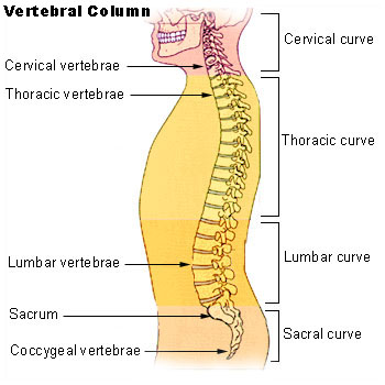
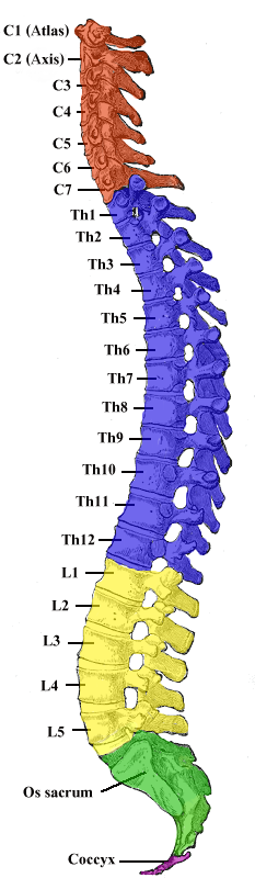
The Vertebral Column:
|
By . - http://training.seer.cancer.gov/module_anatomy/unit3_5_skeleton_divisions.html, Public Domain, https://commons.wikimedia.org/w/index.php?curid=1394201
|
The Spinal Curvature and Vertebral Regions:
By Henry Vandyke Carter - Vertebral column image.- From: Henry Gray (1918) Anatomy of the Human Body (See "Book" section below)- Altered by User:Uwe Gille, Public Domain, https://commons.wikimedia.org/w/index.php?curid=1282158
- C 1-7 (Cervical vertebrae 1-7)
- Neck bones
- C 1 is the atlas and supports the skull
- C 2 is the axis, and rotates the skull (dens is the pivot joint)
- C 3-7 are the smallest and lightest vertebrae
- C 7: vertebra prominens
- Vertebral foramen: slightly triangular
- Spinous process: is short and bifid (2 branches)
- Transverse process: foramina; arteries pass through here to supply the brain
- T 1-12 (Thoracic vertebrae 1-12)
- Middle back
- Larger body, kind of heart-shaped with costal facets on either side
- These articulate with the corresponding ribs
- Vertebral foramen: round or oval
- Spinous process: long, with sharp downward hook
- Forms the posterior part of the rib cage, and are the only vertebrae that articulate with the ribs
- L 1-5 (Lumbar vertebrae 1-5)
- Low back
- Big, cuboidal bodies
- Short, thick spinous processes that extend straight back
- Sturdiest, strongest vertebrae
- Absorb the most shock
- Provide the most support
- Endure the most stress
- L 4-5: between these vertebrae is where spinal taps are performed or where epidurals are inserted
- Sacrum
- A fusion of 5 vertebrae
- Articulates with L 5 and the coccyx
- Median sacral crest: remnant of the spinous processes of the vertebrae that had fused
- Alae: wing-like structures and articulate with the hip bones to form the sacroiliac joints
- Sacral foramina: passageways for blood vessels and nerves
- Sacral canal: vertebral canal continues here
- Sacral hiatus: vertebral column ends here, near the coccyx
- A fusion of 5 vertebrae
- Coccyx (tailbone)
- The fusion of 3-5 small vertebrae
- Remnant of the tail that other vertebrates have
- The fusion of 3-5 small vertebrae
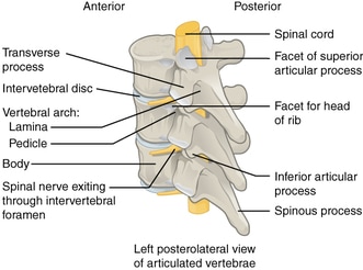
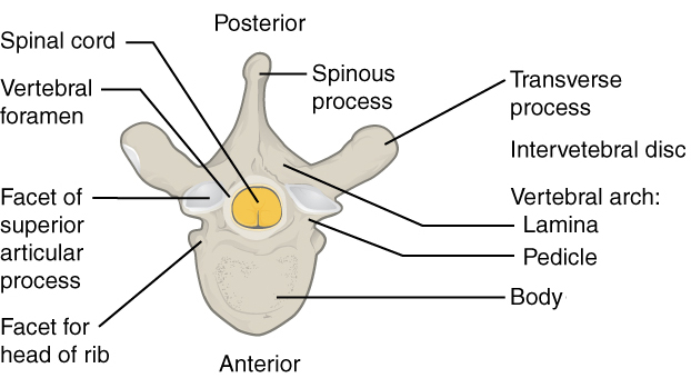
Vertebrae Structure:
|
By Jmarchn - Own work, CC BY-SA 3.0, https://commons.wikimedia.org/w/index.php?curid=45613313
|
By Jmarchn - Own work, CC BY-SA 3.0, https://commons.wikimedia.org/w/index.php?curid=45613314
|
Abnormal Spinal Curvatures:
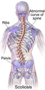
By BruceBlaus. When using this image in external sources it can be cited as:Blausen.com staff (2014). "Medical gallery of Blausen Medical 2014". WikiJournal of Medicine 1 (2). DOI:10.15347/wjm/2014.010. ISSN 2002-4436. - Own work, CC BY 3.0, https://commons.wikimedia.org/w/index.php?curid=27796966
|
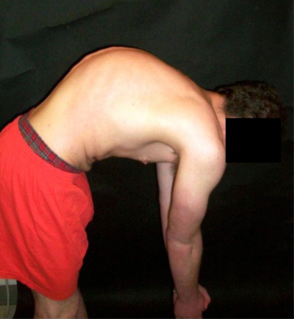
Kyphosis (outward hump);By MusicNewz - Own work this is me, CC0, https://commons.wikimedia.org/w/index.php?curid=15192090
|
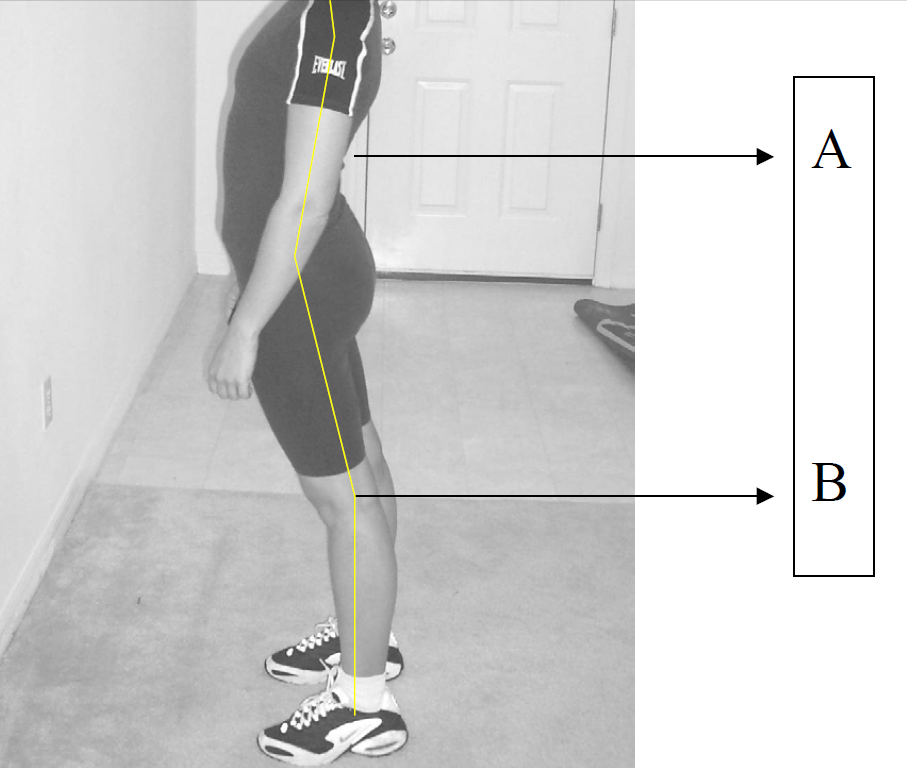
Lordosis; By CarpalTunnelEx - http://www.carpaltunnel-cure.com, CC0, https://commons.wikimedia.org/w/index.php?curid=14165238
|
The Pelvic Girdle:
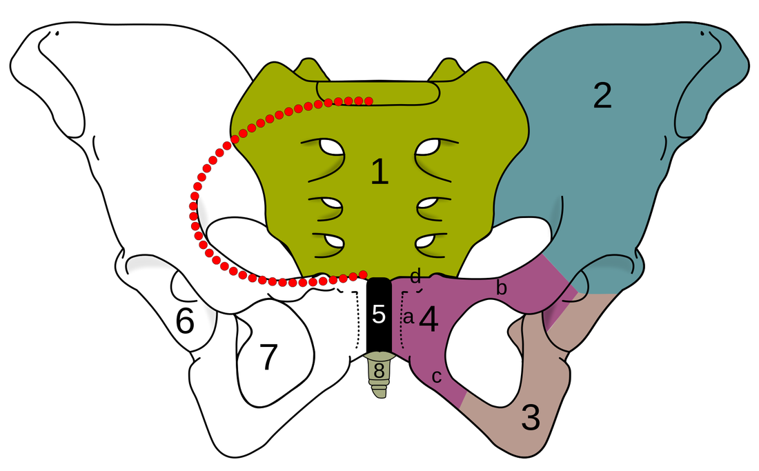
By Fred the Oyster, CC BY-SA 4.0, https://commons.wikimedia.org/w/index.php?curid=35384964
- Sacrum
- Ilium (Iliac Crest), also called the hip bone
- Ischium
- Pubic bone
- a) corpus
- b) ramus superior
- c) ramus inferior
- d) tuberculum pubicum
- Pubic symphysis
- Acetabulum
- Foramen obturatum
- Coccyx (tailbone)
Thorax and Ribs:
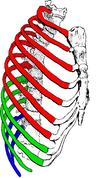
- Fixed Ribs: 1st 7 ribs; attached directly to sternum via cartilage
- False Ribs: Ribs 8, 9, 10, 11, 12; the first 3 sets (8, 9, 10, are attached to cartilage which attaches to cartilage which attaches to sternum)
- Floating Ribs: Ribs 11, 12; not attached to sternum at all
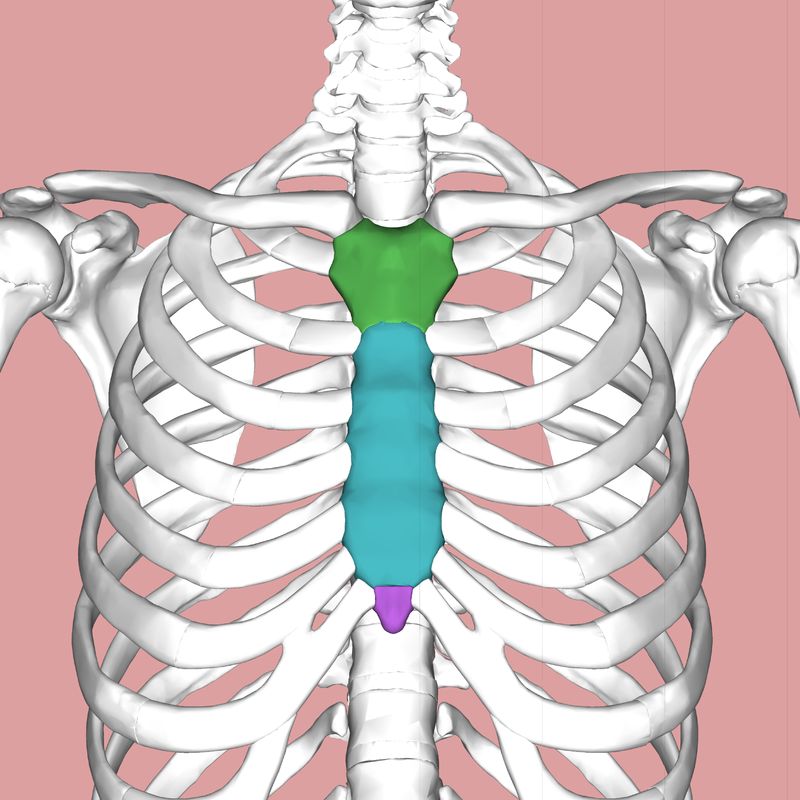
Sternum:
By Anatomography - en:Anatomography (setting page of this image), CC BY-SA 2.1 jp, https://commons.wikimedia.org/w/index.php?curid=24125319
Green: Sternum
Blue: Manubrium
Purple: Xiphoid process
Blue: Manubrium
Purple: Xiphoid process
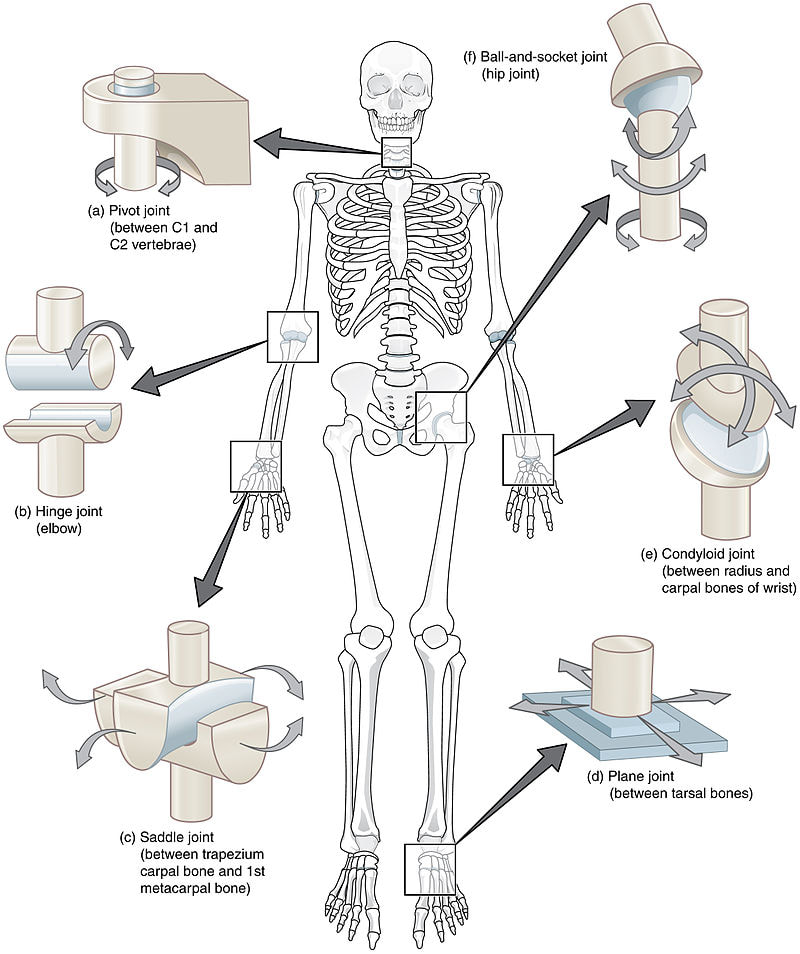
Joints (Articulations):
Structural and Functional Classification of Joints:Learning Objectives:
The Three Major Types of Joints and Their Functions:
- Describe the three types of structural joints and give an example of each.
- Distinguish between the three types of functional joints and give an example of each.
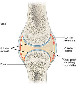
- Describe the basic structure of a typical synovial joint.
- Describe the types of movements of synovial joints and demonstrate them.
The Three Major Types of Joints and Their Functions:
- Fibrous joints - immovable joints or synarthroses (sutures between skull bones, teeth sockets)
- Cartilaginous joints - slightly moveable joints or amphiarthroses (intervertebral joints, tibiofibular joint, pubic symphysis)
- Synovial joints - fully moveable joints or diarthroses (about 90% of the joints in the body) (knee, shoulder, hip, elbow)