The Human Digestive System:
Lecture Objective: Chapters 16-17 Digestive System:Upon completion of this module, you should be able to:
Lab Objectives: Exercises 21-22, Structure of the Digestive System and Mechanical and Chemical Digestion:Upon completion of these exercises, you should be able to:
- List and describe the four layers of the wall of the alimenatary canal.
- Compare the lining of the esophagus, stomach, small intestine, and large intestine.
- Define and contrast the mechanical and chemical digestion processes.
- Discuss the basics of carbohydrate, protein, and fat digestion and give the end products of each process.
- List the sequence each of the component parts of segments of the alimentary canal from the mouth to the anus.
- Identify the accessory organs of digestion.
- Define the terms peristalsis, bolus, chyme, jaundice, ulcer and diarrhea.
- Describe some of the digestive system disorders, diseases and conditions.
- Explain metabolism and compare and contrast anabolism and catabolism.
- Explain the structure and function of an enzyme and its role in digestion.
- Describe the metabolic roles of carbohydrates, lipids, proteins, vitamins, and minerals.
- Define basal metabolic rate (BMR), and list some factors that can affect it.
- Discuss the physiological mechanisms that regulate body temperature.
Lab Objectives: Exercises 21-22, Structure of the Digestive System and Mechanical and Chemical Digestion:Upon completion of these exercises, you should be able to:
- Identify the major gastrointestinal (GI) tract organs and accessory digestive organs on charts and models.
- Describe the peritoneum and identify the major peritoneal structures.
- Describe the functions of each GI tract organ and accessory digestive organ.
- Name the substrate for each of the following enzymes and the products produced from the enzyme-catalyzed reaction: amylase, protease, lipase, and peptidase.
- Describe the importance of mechanical and chemical digestion.
- Describe the importance of pH on digestive enzyme function.
- Describe the action of bile salts on lipids.
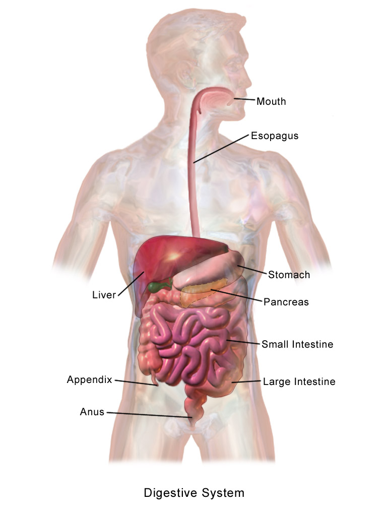
The human digestive system is very complex and includes many structures and organs. Digestion begins at the mouth and includes all structures in the mouth, saliva, digestive enzymes present in saliva, and includes the esophagus, stomach, gallbladder, pancreas, liver, small intestine, large intestine, appendix, anus and rectum. Some of these organs are found in other organ systems as well.
Digestive system - contains the organs of the GI tract and accessory digestive organs: the mouth, most of the pharynx, esophagus, stomach, small intestine, large intestine, anus, salivary glands, tongue, teeth, pancreas, liver, and gallbladder.
GI Tract - is the gastrointestinal tract, also known as the alimentary tract or canal, a tube extending from the mouth to the anus.
Lumen - the space of the GI tract that opens to the external environment at either end.
GI Tract - is the gastrointestinal tract, also known as the alimentary tract or canal, a tube extending from the mouth to the anus.
Lumen - the space of the GI tract that opens to the external environment at either end.
By BruceBlaus - Own work, CC BY-SA 4.0, https://commons.wikimedia.org/w/index.php?curid=57309988
The Peritoneum:Peritoneum - this is the largest serous membrane in the body, and is located in the abdominopelvic cavity. It is a visceral and a parietal layer.
Parietal peritoneum - lines the inner surface of the abdominopelvic wall.
Visceral peritoneum - covers the organs within the abdominopelvic cavity. It is also called the serosa.
Peritoneal (serous) fluid - fluid found between the 2 peritoneal layers.
Peritoneal cavity - space between the 2 peritoneal layers.
Retroperitoneal organs - organs that lie outside the peritoneal cavity.
Falciform ligament - large folds not found in other serous membranes that secure organs together and to the abdominal walls. This one binds the liver to the anterior abdominal wall.
Greater omentum - large folds not found in other serous membranes that secure organs together and to the abdominal walls. This one is a large, fatty-looking apron that folds back on itself and attaches to the transverse colon, forming a pouch that hangs between the body wall and the anterior surface of the small intestine.
Mesentery - part of the peritoneum that holds the small intestine to the posterior abdominal wall.
Gastrointestinal Tract Organs: The Mouth:
Mouth - food first enters here, and the mouth is called the oral or buccal cavity proper.
Hard palate - the palatine bone and palatine processes of the maxillae that form the hard roof of the mouth.
Soft palate - forms the roof of the mouth and is muscular, forming the two arches that border the fauces, and hangs down to form the oval process called the uvula. During swallowing, this structure keeps food from entering the nasal cavity.
Tongue - forms the floor of the mouth
Cheeks - form the lateral walls of the mouth.
Lips - form the anterior border of the mouth.
Fauces - form the posterior border of the mouth and opening to the oropharynx.
Palatine tonsils - are located at the back of the throat between the two arches of the soft palate with the uvula hanging down in between.
The Pharynx:
Pharynx - the throat:
Epiglottis - part of the larynx that closes off the inferior portion of the laryngophraynx during swallowing, allowing food and drink to enter the esophagus.
Mouth - food first enters here, and the mouth is called the oral or buccal cavity proper.
Hard palate - the palatine bone and palatine processes of the maxillae that form the hard roof of the mouth.
Soft palate - forms the roof of the mouth and is muscular, forming the two arches that border the fauces, and hangs down to form the oval process called the uvula. During swallowing, this structure keeps food from entering the nasal cavity.
Tongue - forms the floor of the mouth
Cheeks - form the lateral walls of the mouth.
Lips - form the anterior border of the mouth.
Fauces - form the posterior border of the mouth and opening to the oropharynx.
Palatine tonsils - are located at the back of the throat between the two arches of the soft palate with the uvula hanging down in between.
The Pharynx:
Pharynx - the throat:
- Nasopharynx - the superior area, posterior and inferior to the nose, which has respiratory functions.
- Oropharynx - the middle area, posterior to the fauces of the oral cavity, which has both respiratory and digestive functions because air, food and liquid pass through it.
- Laryngopharynx - the inferior area located posterior to the larynx, which is also a common passageway for air, food and liquid.
Epiglottis - part of the larynx that closes off the inferior portion of the laryngophraynx during swallowing, allowing food and drink to enter the esophagus.
Esophagus and Stomach:
Esophagus - the tube that is posterior to the trachea, in which food and drink move from the laryngopharynx to the superior part of the stomach.
Stomach - J-shaped pouch lying against the diaphragm superiorly, which has four areas: the cardia, the fundus, the body and the pylorus.
Cardia - first part of the stomach, inferior to the esophagus.
Fundus - the elevated area to the left of the cardia that serves as a temporary holding area for food.
Body - the largest, curved part of the stomach.
Pylorus - funnel-shaped region of the stomach, continuing to the right of the body of the stomach after the curve.
Pyloric sphincter - the smooth muscle between the stomach and small intestine that allows food to enter the first part of the small intestine, the duodenum.
Rugae - the large, conspicuous ridges in the mucosa that allow the stomach to be stretched when food and drink are present and which disappear as the stomach becomes fully distended.
Small Intestine and Large Intestine:
Small Intestine:
Small intestine - is a long tube that is about ten feet in length, which is divided into three sections: the duodenum, the jejunum, and the ileum. This is the major site of digestion and mineral absorption.
Duodenum - shortest part of the small intestine, first section that is directly attached to the stomach, is shaped like a "C", and receives food through the pyloric sphincter. It is a little less than 1' long.
Jejunum - the middle section of the small intestine, following the duodenum, which is about 3' long and has a thicker layer of smooth muscle. It is empty upon death.
Ileum - the final section of the small intestine, about 6' in length, which ends at the ileocecal sphincter (valve) where it joins the large intestine.
Large Intestine:
Large intestine - the large bowel, or colon, which is around 5' long, is composed of the cecum, colon, rectum, and anal canal, and absorbs a large amount of water, concentrating the undigested feces into a solid form.
Cecum - blind pouch about 2-3" long that extends inferiorly from the large intestine at the ileocecal sphincter (valve) in the lower right quadrant.
Appendix - vermiform appendix branches off the cecum and is about 2-3" long.
Ascending colon - extends superiorly to the liver on the right side of the abdomen and makes a 90 degree turn to the left.
Transverse colon - the part of the colon that lies straight across.
Descending colon - continues inferiorly on the left side to an S-shaped curve.
Sigmoid colon - S-shaped curve following the descending colon, found at the level of the iliac crest.
Haustra - bands of smooth muscle on the exterior of the colon that contract to form many pouches, giving it a gathered appearance.
Rectum - follows the sigmoid colon, at about the level of the 3rd sacral vertebrae.
Anal canal - last segment of the colon, opening to the exterior
Anus - portion of the anal canal that opens to the exterior for excretion, containing 2 sets of sphincters that control elimination of feces (internal and external sphincters).
Sigmoidoscopy - a scope with a camera that views the lining of the sigmoid colon.
Colonoscopy - a scope with a camera that views the lining of the entire colon.
Accessory Digestive Organs:
Salivary Glands:
Mucous Membranes - the mucous membranes of the mouth and tongue have many small salivary glands that secrete saliva.
Salivary Glands - most of the saliva secreted into the oral cavity is from the parotid, submandibular, and sublingual salivary glands.
Parotid Gland - largest salivary gland, which is located anterior and inferior to the ears to the ears between the skin and the masseter muscle.
Submandibular Glands - located in the posterior part of the mouth floor just medial to the mandible.
Sublingual Glands - located under the tongue as the name suggests, more medial and superior than the submandibular glands.
Tongue:
Extrinsic muscles - these muscles hold the tongue in position and allow movement to maneuver food in the mouth.
Intrinsic muscles - the tongue is composed of this type of skeletal muscle, which is used for speech and swallowing.
Lingual frenulum - attaches the tongue to the floor of the mouth.
Teeth:
Teeth - mechanically break up food into small pieces and area aided by the tongue that manipulates food in the oral cavity.
The Pancreas, Liver and Gallbladder:
Pancreas:
Pancreas - is located posterior to the stomach and lies in the C-shape of the duodenum and secretes digestive enzymes that travel to the duodenum through the pancreatic duct.
Pancreatic duct - a duct that joins the common bile duct to empty into the duodenum.
Liver:
Liver - a large organ that lies inferior to the diaphragm and touches the diaphgram along the liver's superior border, and has two main lobes: a right lobe and a left lobe, and makes the bile that is delivered to the gallbladder for concentration and storage.
Falciform ligament - separates the liver into two main lobes, the right lobe and the left lobe.
Common hepatic duct - carries bile to the cystic duct of the gallbladder.
Gallbladder:
Gallbladder - a sac made of smooth muscle that lies on the inferior front part of the liver.
Cystic duct - bile leaves the gallbladder from here and joins with the common hepatic duct to form the common bile duct.
Common bile duct - joins the pancreatic duct to enter the duodenum, and it is at this site that bile salts aid in transforming large fat globules into smaller fat particles for rapid digestion to occur by catabolism.
Mechanical And Chemical Digestion, Lab Exercise 27:Chemical Digestion:
Macromolecules - large molecules contained in the food we eat, including carbohydrates, proteins, and lipids. Examples:
Enzymes - biological catalysts that kick off and speed up the rate of chemical reactions and metabolism, in order to break down large macromolecules into smaller ones that can be absorbed. They act on substrates to produce products.
Catabolism - the breakdown of macromolecules into monomers or monosaccharides, glycerol, fatty acids, and amino acids by enzymes, which are then small enough to be absorbed across the wall of the gastrointestinal tract and are used by cells to build new macromolecules or to provide energy (ATP).
Anabolism - the building of new larger macromolecules from smaller monomers.
Macromolecules - large molecules contained in the food we eat, including carbohydrates, proteins, and lipids. Examples:
- Starch - a polysaccharide (complex) carbohydrate that is digested into disaccharides (lactose, sucrose, maltose) and into monosaccharides (glucose, fructose, galactose) by the action of specific enzymes called amylases.
- Lipids - macromolecules that are catabolized or digested into glycerol and fatty acids by the enzyme lipase.
- Proteins - macromolecules that are catabolized or digested into peptides and amino acids by the enzyme protease. Peptides are then further digested by peptidases into amino acids.
Enzymes - biological catalysts that kick off and speed up the rate of chemical reactions and metabolism, in order to break down large macromolecules into smaller ones that can be absorbed. They act on substrates to produce products.
- They work best on food substrates that have been chewed up and broken down mechanically into smaller particles with a greater surface area.
- Each has an optimal environmental condition, including temperature and pH, that allow it to be the most active.
- Enzymes in our body function well at body temperature (37 degrees C or 98.6 degrees F)
- Enzymes become denatured and inactive if the temperature becomes too high.
- The pH of the digestive system changes from the mouth to the small intestine, so different areas have different optimal pH's.
Catabolism - the breakdown of macromolecules into monomers or monosaccharides, glycerol, fatty acids, and amino acids by enzymes, which are then small enough to be absorbed across the wall of the gastrointestinal tract and are used by cells to build new macromolecules or to provide energy (ATP).
Anabolism - the building of new larger macromolecules from smaller monomers.
Example of a Major Specific Digestive Enzyme: Amylase:
Salivary amylase - digests carbohydrates (starch) into glucose (a monosaccharide).
When we eat starch (cracker, cereal, bread, potato, etc...) it is:
Salivary amylase - digests carbohydrates (starch) into glucose (a monosaccharide).
When we eat starch (cracker, cereal, bread, potato, etc...) it is:
- Mechnically broken down and digested with the teeth and tongue.
- Chemically broken down by the enzyme amylase.
- Carbohydrate digestion begins in the mouth, at a pH of 7.4.
- Salivary amylase is denatured by the low pH in the stomach from the presence of hydrochloric acid (HCl).
- Pancreatic buffers bring the pH back to 7.4, favorable for pancreatic amylase, allowing carbohydrate digestion to continue in the small intestine.
Using the Images Below, Read Activity 1, Digestion of Starch, Then Complete Table 27.1 and Discussion Questions: Digestion of Starch, Activity 2: Bile Emulsification and Questions and Discussion Questions, and Reviewing Your Knowledge:
Starch Test- Lugol's Iodine (IKI) is used as a color indicator to indicate the presence of starch. Lugol's iodine is tea-colored on its own. When starch is present, the sample will turn dark blue-to-purple-to-black, depending upon the amount of starch present, indicating a positive starch test. A negative starch test remains tea-color.
Starch Test- Lugol's Iodine (IKI) is used as a color indicator to indicate the presence of starch. Lugol's iodine is tea-colored on its own. When starch is present, the sample will turn dark blue-to-purple-to-black, depending upon the amount of starch present, indicating a positive starch test. A negative starch test remains tea-color.
- Light blue (+)
- Medium blue (++)
- Dark blue/purple (+++)
- Black (++++)
The Benedict's Test for Sugar:
Benedict's solution - this is a light blue copper solution that changes colors if a reducing sugar is present, indicating a positive sugar test.
Benedict's solution - this is a light blue copper solution that changes colors if a reducing sugar is present, indicating a positive sugar test.
- A positive sugar test - this will range in color from green to yellow to orange to red to brick-red, depending upon the amount of sugar present.
- Green (+)
- Yellow (++)
- Orange (+++)
- Red (++++)
- A negative sugar test - will stay light blue and there will be no color change.
- Blue (-)
How do you think that the results of the whole piece of cracker differ from one that has been crumbled or chewed up?
When a cracker is chewed or crumbled, it has been mechanically and even chemically digested, increasing the surface area for amylase to work on and speed up the reaction. A whole cracker produces less sugar since there is a decreased surface area for amylase to work on.
Amylase enzyme works at the ideal pH of 7.4, which is the pH of the mouth and saliva in the mouth. An acidic pH in the HCl of the stomach denatures it and breaks it down, making the enzyme inactive.
In the activity, this is how the tubes would be set up:
Tubes 3 and 4 are positive for starch.
Tubes 3 and 4 are positive for sugar.
None of the tubes were positive just for sugar. Not all the starch was broken down into sugar, so ALL tubes were positive for starch.
A longer incubation time at the right temperature of 37 degrees C means that more sugar would appear in tubes with amylase at a pH of 7.4, since heat increases the enzyme activity.
Tube 4 is similiar to the conditions of the stomach, because it contained the crumbled cracker, amylase, and HCl acid. The stomach receives broken down food from the mouth that was mechanically and chemically broken down by the teeth and amylase, going to the acidic HCl acid of the stomach, where amylase is denatured by the acid.
Bile Emulsification:
Bile - composed of a mix of substances, including bile salts, that aid in digestion by physically breaking up large molecules of lipids into smaller molecules (glycerol and fatty acids) with a greater surface area. Bile is not an enzyme. It is an emulsifier.
When a cracker is chewed or crumbled, it has been mechanically and even chemically digested, increasing the surface area for amylase to work on and speed up the reaction. A whole cracker produces less sugar since there is a decreased surface area for amylase to work on.
Amylase enzyme works at the ideal pH of 7.4, which is the pH of the mouth and saliva in the mouth. An acidic pH in the HCl of the stomach denatures it and breaks it down, making the enzyme inactive.
In the activity, this is how the tubes would be set up:
- Tube 1: Control (whole cracker, buffer, water, iodine, Benedict's solution)
- Tube 2: Control (crumbled cracker, buffer, water, iodine, Benedict's solution)
- Tube 3: Whole cracker, buffer, amylase, water, iodine, Benedict's solution
- Tube 4: Crumbled cracker, buffer, amylase, water, iodine, Benedict's solution
- Tube 5: Whole cracker, buffer, amylase, 0.5% HCl acid, iodine, Benedict's solution
- Tube 6: Crumbled cracker, buffer, amylase, 0.5% HCl acid, iodine, Benedict's solution
Tubes 3 and 4 are positive for starch.
Tubes 3 and 4 are positive for sugar.
None of the tubes were positive just for sugar. Not all the starch was broken down into sugar, so ALL tubes were positive for starch.
A longer incubation time at the right temperature of 37 degrees C means that more sugar would appear in tubes with amylase at a pH of 7.4, since heat increases the enzyme activity.
Tube 4 is similiar to the conditions of the stomach, because it contained the crumbled cracker, amylase, and HCl acid. The stomach receives broken down food from the mouth that was mechanically and chemically broken down by the teeth and amylase, going to the acidic HCl acid of the stomach, where amylase is denatured by the acid.
Bile Emulsification:
Bile - composed of a mix of substances, including bile salts, that aid in digestion by physically breaking up large molecules of lipids into smaller molecules (glycerol and fatty acids) with a greater surface area. Bile is not an enzyme. It is an emulsifier.
- Made by the liver
- Stored in the gallbladder
