The Urinary System and the Urinalysis:
Lecture Objectives, Chapter 18: Urinary SystemUpon completion of this chapter, you should be able to:
Lab Objectives, Exercise 28: Structure of the Urinary System and Urinalysis and Sheep Kidney DissectionUpon completing these exercises, you should be able to:
- Identify the major organs of the urinary system and give the generalized function of each.
- Name the parts of a nephron and describe the role each component plays in the formation of urine.
- Explain how the kidneys act as vital organs in maintaining homeostasis.
- Explain the importance of filtration, reabsorption, and secretion in urine formation.
- Discuss the mechanisms that control urine volume, including the normal amount and composition of urine.
- Explain the process of urine elimination.
Lab Objectives, Exercise 28: Structure of the Urinary System and Urinalysis and Sheep Kidney DissectionUpon completing these exercises, you should be able to:
- Locate and identify the organs of the urinary system on models or charts.
- Describe how urine is formed.
- Trace the path of urine from formation through the path it takes to the exterior of the body.
- Describe the major features of a urinalysis and its purpose and components.
- Dissect a sheep kidney, identifying the major features of the kidney.
Urinary System:
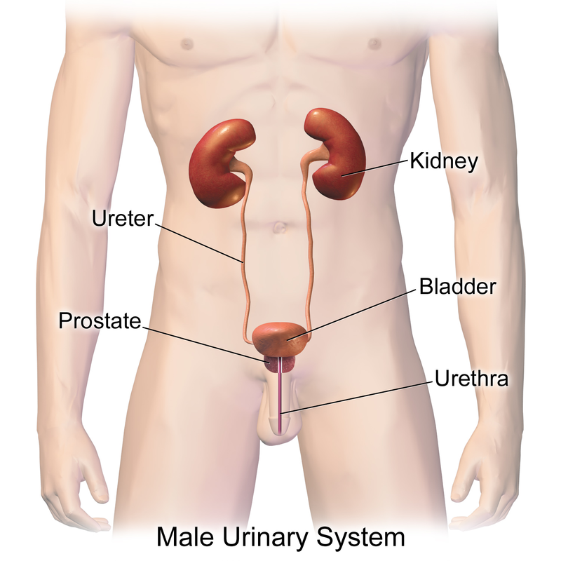
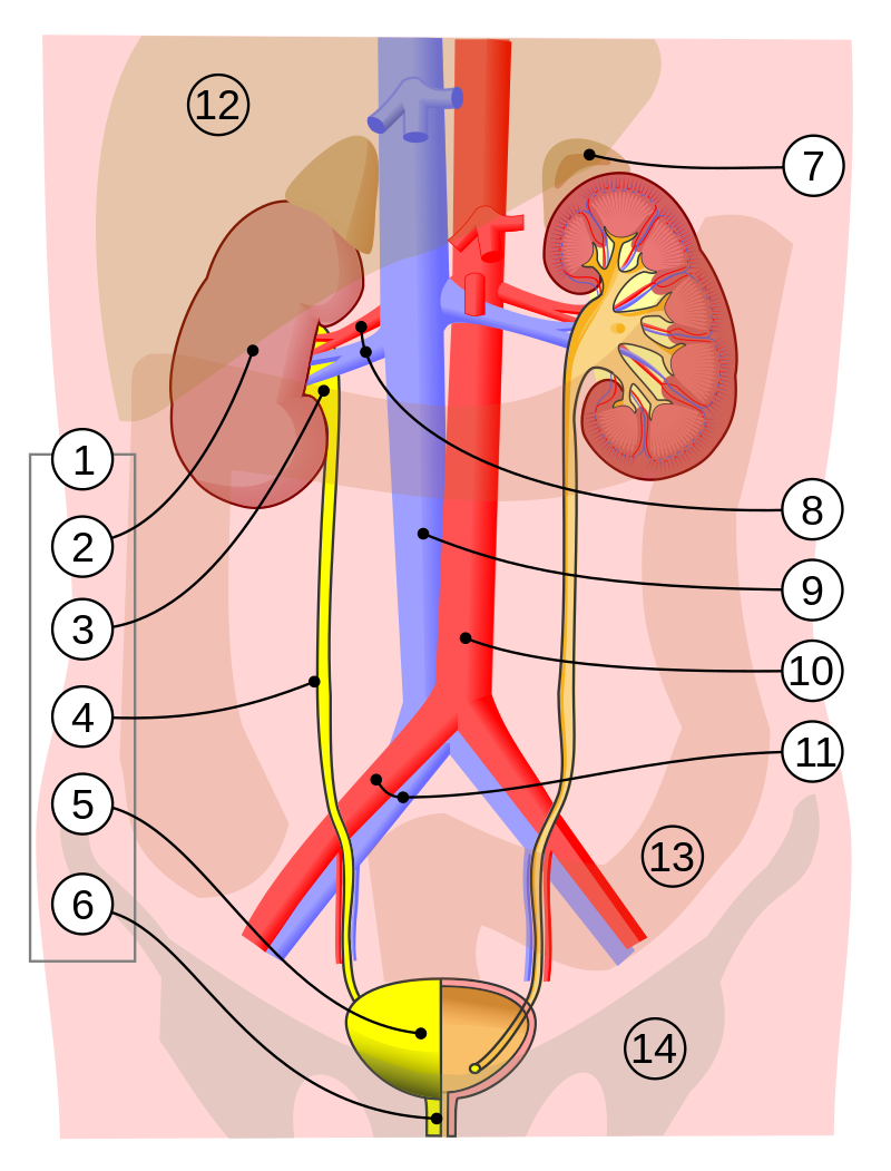
Urinary System Organs: include the kidneys, ureters, urinary bladder, and urethra.
Kidney - regulates water, electrolyte, and pH balance in the body. It removes wastes and nitrogenous substances from blood and excretes them in the urine. They are bean-shaped structures that sit retroperitoneal between the abdominal wall and the peritoneum, are found at the waste level between the 12th thoracic vertebra and the 3rd lumbar vertebra.
Urine - formed in the nephrons of the kidneys and flows through the ureters to the urinary bladder that stores urine until it is eliminated from the body through the urethra.
Retroperitoneal - sitting behind
Renal - pertaining to the kidneys
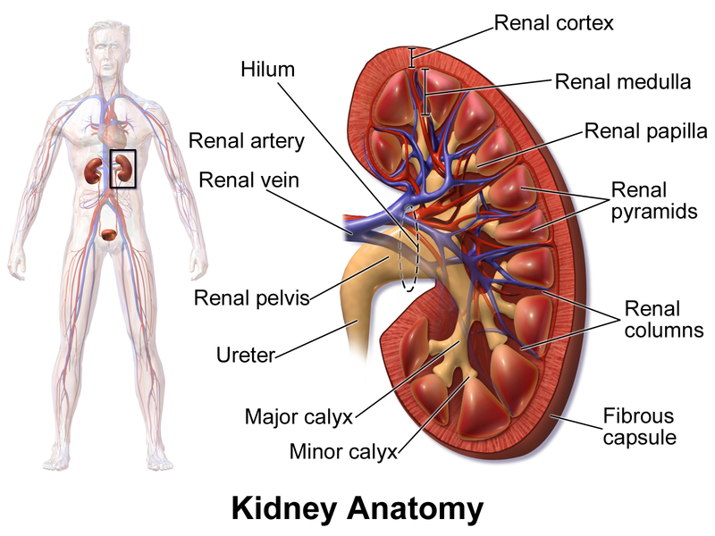
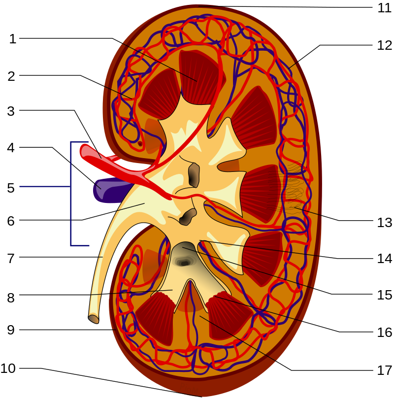
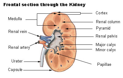
Renal capsule - thin, fibrous membrane that covers the outer surface of the kidney
Renal cortex - the most superficial region of the kidney
Renal medulla - deep to the cortex
Renal pyramids - cone-shaped structures found in the medulla
Renal columns - extensions of the cortex found between the renal pyramids
Base - bottom of each pyramid that faces the cortex
Renal papilla - the apex that is pointed toward the center of the kidney
Nephrons - found in the medulla and cortex, and are the structural and functional units of the kidneys that form urine
Calyces (calyx, singular) - major and minor ducts that exit through openings of the renal papilla and are cup-shaped and carry urine to the renal pelvis
Renal pelvis - urine drains here from the calyces, and it connects to the ureter
Ureters - tubes that drain urine from the renal pelvis to the urinary bladder
Complete Activity 1: Location and Structure of the Kidneys
Urinary System Organs: include the kidneys, ureters, urinary bladder, and urethra.
Kidney - regulates water, electrolyte, and pH balance in the body. It removes wastes and nitrogenous substances from blood and excretes them in the urine. They are bean-shaped structures that sit retroperitoneal between the abdominal wall and the peritoneum, are found at the waste level between the 12th thoracic vertebra and the 3rd lumbar vertebra.
- The right kidney is slightly lower than the left kidney
- The liver forces the right kidney into a lower position
Urine - formed in the nephrons of the kidneys and flows through the ureters to the urinary bladder that stores urine until it is eliminated from the body through the urethra.
Retroperitoneal - sitting behind
Renal - pertaining to the kidneys
Renal capsule - thin, fibrous membrane that covers the outer surface of the kidney
Renal cortex - the most superficial region of the kidney
Renal medulla - deep to the cortex
Renal pyramids - cone-shaped structures found in the medulla
Renal columns - extensions of the cortex found between the renal pyramids
Base - bottom of each pyramid that faces the cortex
Renal papilla - the apex that is pointed toward the center of the kidney
Nephrons - found in the medulla and cortex, and are the structural and functional units of the kidneys that form urine
Calyces (calyx, singular) - major and minor ducts that exit through openings of the renal papilla and are cup-shaped and carry urine to the renal pelvis
Renal pelvis - urine drains here from the calyces, and it connects to the ureter
Ureters - tubes that drain urine from the renal pelvis to the urinary bladder
Complete Activity 1: Location and Structure of the Kidneys
The Ureters, Urinary Bladder and Urethra:
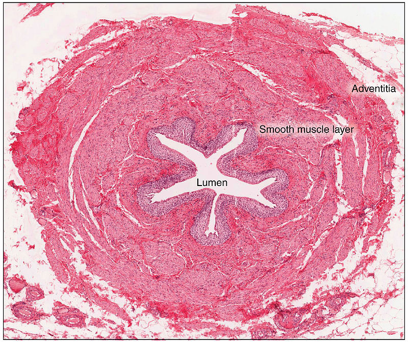
Ureters - narrow, long muscular tubes also located in a retroperitoneal position, descending toward the urinary bladder, curving medially, as they approach the inferior portion of the bladder, and enter the posterior wall of the bladder at an oblique angle.
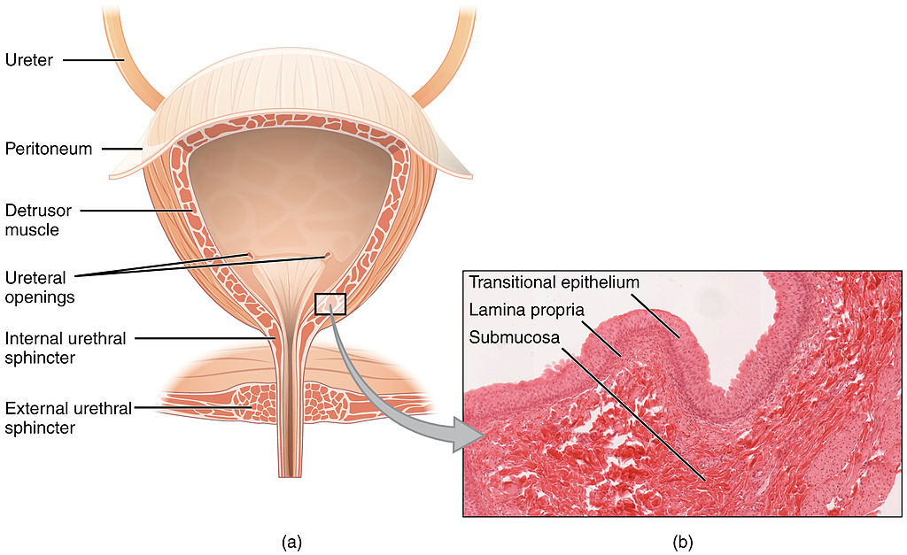
Urinary bladder - a hollow, muscular organ that distends to store urine and has three internal openings that form a triangle called the trigone.
Micturition - urination or voiding.
Internal urethral sphincter - a layer of circular, involuntary smooth muscle that controls passage of urine into the urethra from the urinary bladder.
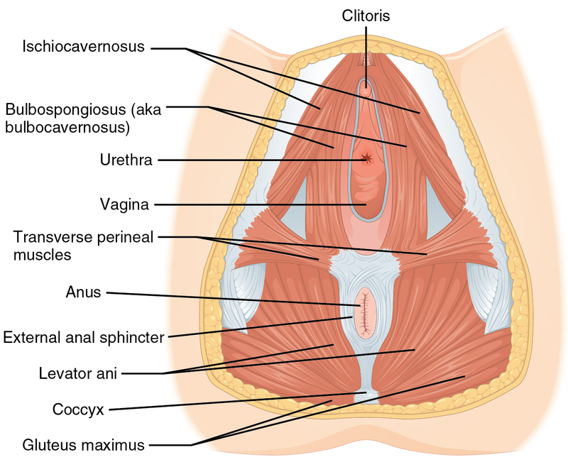
External urethral sphincter - voluntary skeletal muscle within the urogenital diaphragm (deep muscles of the perineum) that permit the passage of urine to the external urethral orifice.
Benign prostatic hypertrophy - a noncancerous enlargement of the prostate gland that restricts the urethra and inhibits urine flow, causing urinary retention.
Ureters - narrow, long muscular tubes also located in a retroperitoneal position, descending toward the urinary bladder, curving medially, as they approach the inferior portion of the bladder, and enter the posterior wall of the bladder at an oblique angle.
Urinary bladder - a hollow, muscular organ that distends to store urine and has three internal openings that form a triangle called the trigone.
- The 2 posterior openings are the ureters.
- The anterior opening is the internal urethral orifice into the urethra.
Micturition - urination or voiding.
Internal urethral sphincter - a layer of circular, involuntary smooth muscle that controls passage of urine into the urethra from the urinary bladder.
External urethral sphincter - voluntary skeletal muscle within the urogenital diaphragm (deep muscles of the perineum) that permit the passage of urine to the external urethral orifice.
Benign prostatic hypertrophy - a noncancerous enlargement of the prostate gland that restricts the urethra and inhibits urine flow, causing urinary retention.
A urinalysis is a group of tests performed to see whether or not the urine is normal or abnormal. Urine can tell a lot about someone's health and underlying conditions, from dehydration, to inability to concentrate the urine, to diabetes, to ketoacidosis, to various types of infections, and much more. A urinalysis panel of tests includes:
- Collection of urine following special protocol, making sure it is properly collected and labeled, making sure it is sealed properly, and sending it to the lab within 2 hours of collection
- Properly preserving the urine until it can be examined
- Macroscopic analysis: color, odor, turbidity, amount/volume, any blood present, dye present, lipids floating, or even kidney stones
- Microscopic analysis: looking at the urine (following concentration of urine) sediment for the presence of any cells (epithelial, red blood cells, white blood cells/leukocytes), bacteria, yeasts, protists, casts, or crystals
- Multistix dipstix, which test for the presence of glucose, protein, lipids, ketones, bilirubin, urobilinogen, blood, leukocytes, nitrites, and specific gravity
- Specific gravity (can be determined by a urine hydrometer or a test strip or the IRIS automated analyzer)
Urinary System:
https://upload.wikimedia.org/wikipedia/commons/thumb/6/6d/Urinary_System_%28Male%29.png/800px-Urinary_System_%28Male%29.png
|
https://upload.wikimedia.org/wikipedia/commons/thumb/d/d6/Blausen_0592_KidneyAnatomy_01.png/800px-Blausen_0592_KidneyAnatomy_01.png
|
https://upload.wikimedia.org/wikipedia/commons/thumb/a/ab/KidneyStructures_PioM.svg/800px-KidneyStructures_PioM.svg.png
|
|
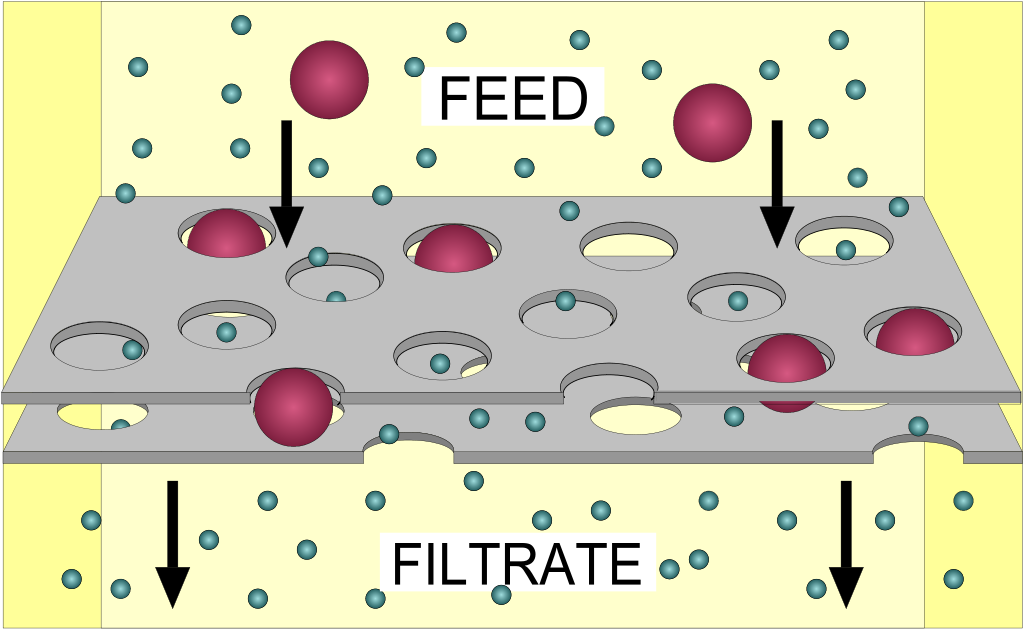
https://upload.wikimedia.org/wikipedia/commons/thumb/2/20/FilterDiagram.svg/1024px-FilterDiagram.svg.png
https://upload.wikimedia.org/wikipedia/commons/thumb/9/93/Hemodialysis-en.svg/654px-Hemodialysis-en.svg.png
https://upload.wikimedia.org/wikipedia/commons/thumb/a/ae/Osmosis_Diffusion_Ultrafiltration_and_Dialysis.svg/800px-Osmosis_Diffusion_Ultrafiltration_and_Dialysis.svg.png
|
https://upload.wikimedia.org/wikipedia/commons/thumb/f/fe/Semipermeable_membrane_%28svg%29.svg/800px-Semipermeable_membrane_%28svg%29.svg.png
|
https://upload.wikimedia.org/wikipedia/commons/thumb/2/2b/Physiology_of_Nephron.png/800px-Physiology_of_Nephron.png
https://upload.wikimedia.org/wikipedia/commons/thumb/6/69/Renal_corpuscle-en.svg/1024px-Renal_corpuscle-en.svg.png
https://upload.wikimedia.org/wikipedia/commons/thumb/2/28/Juxtaglomerular_Apparatus_and_Glomerulus.jpg/1920px-Juxtaglomerular_Apparatus_and_Glomerulus.jpg
https://upload.wikimedia.org/wikipedia/commons/thumb/2/2e/2611_Blood_Flow_in_the_Nephron.jpg/800px-2611_Blood_Flow_in_the_Nephron.jpg
https://upload.wikimedia.org/wikipedia/commons/thumb/3/30/Urinary_system.svg/800px-Urinary_system.svg.png
https://upload.wikimedia.org/wikipedia/commons/thumb/1/17/2607_Ureter.jpg/800px-2607_Ureter.jpg
|
https://upload.wikimedia.org/wikipedia/commons/5/56/Ultrasound_demonstration_of_ureteral_jet_effect_0301105703_1105300.gif
|
https://upload.wikimedia.org/wikipedia/commons/thumb/d/dc/2605_The_Bladder.jpg/1024px-2605_The_Bladder.jpg
https://upload.wikimedia.org/wikipedia/commons/thumb/f/f9/1116_Muscle_of_the_Female_Perineum.png/800px-1116_Muscle_of_the_Female_Perineum.png
Urinalysis:
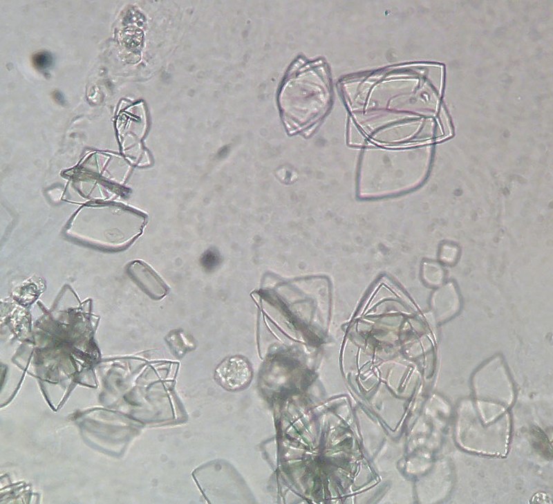
Uric acid crystals
|
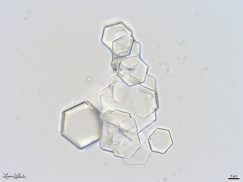
Cystine Crystals
|
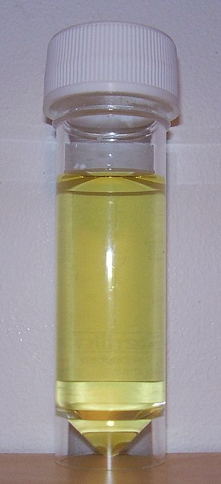
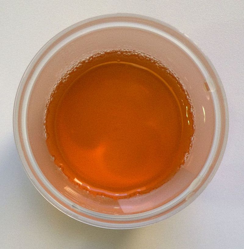
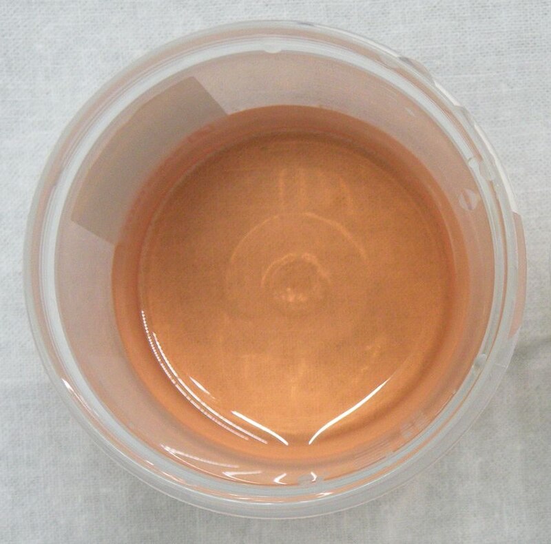
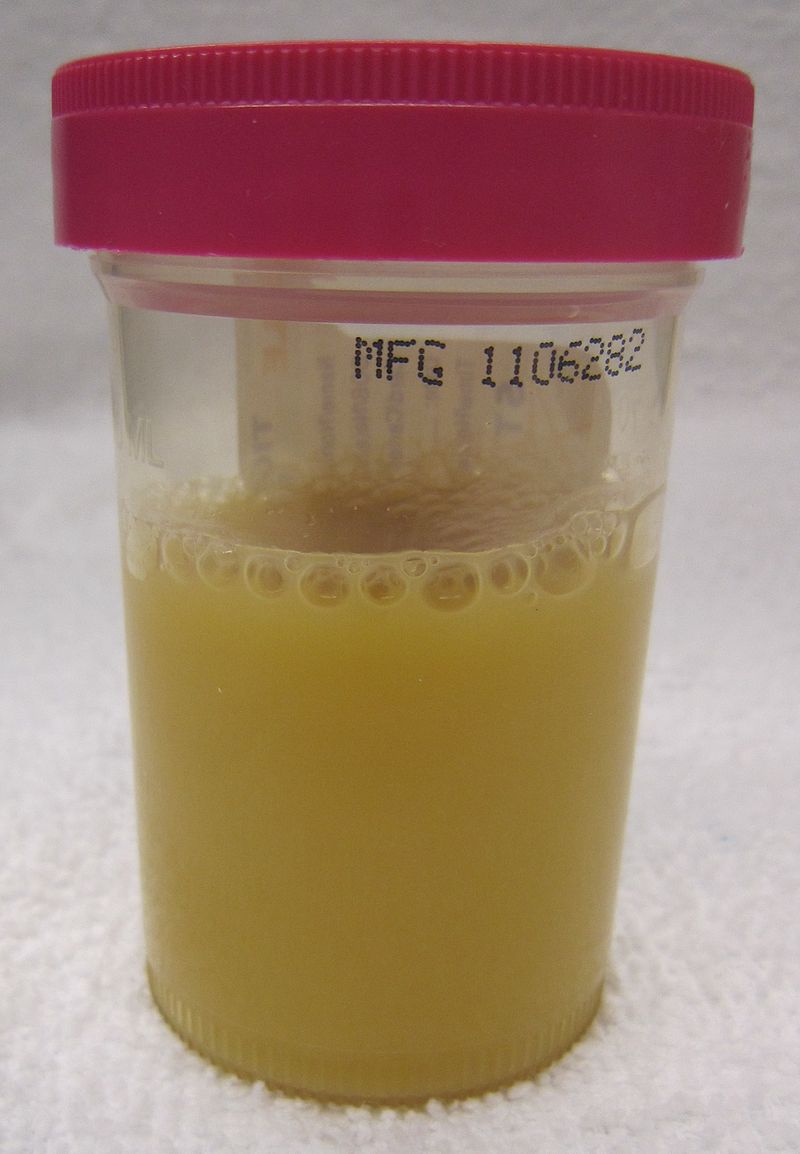
Pale straw, normal color of urine, is seen here. Urine is mostly water, with dissolved minerals, vitamins, waste products, proteins, and should be free of microbes, cells, and a pH of about 6.4 on average.
|
|
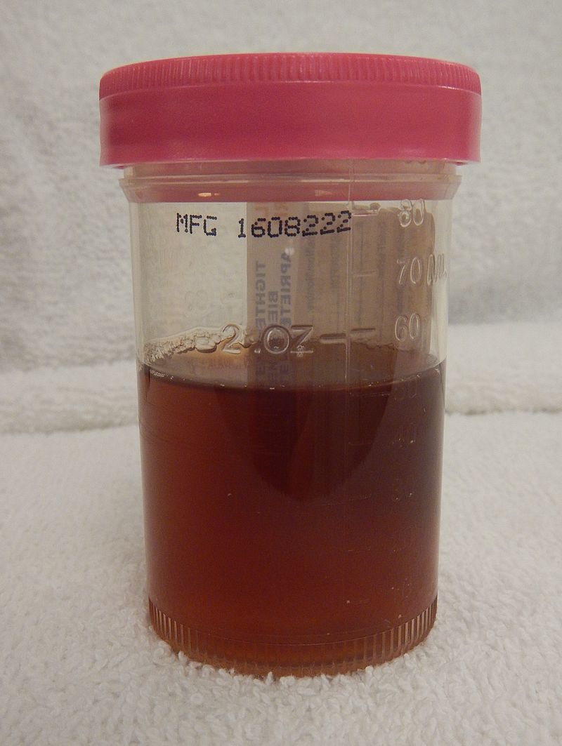
Dark urine due to dehydration, https://upload.wikimedia.org/wikipedia/commons/thumb/6/6a/Dark_urine_due_low_fluid_intake.jpg/800px-Dark_urine_due_low_fluid_intake.jpg
|
Hematuria, https://upload.wikimedia.org/wikipedia/commons/thumb/e/e9/HematuriaGross.jpg/800px-HematuriaGross.jpg
|
Choluria (gallstones), https://upload.wikimedia.org/wikipedia/commons/thumb/2/2c/Choluria.svg/800px-Choluria.svg.png
|
Post-consumption of beets, https://upload.wikimedia.org/wikipedia/commons/thumb/d/df/Pinkish_urine_beetroots_1.jpg/800px-Pinkish_urine_beetroots_1.jpg
|
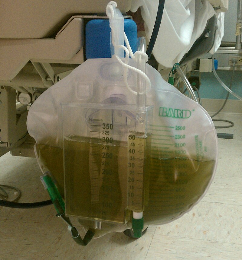
Propofol sedative is one thing that can turn the urine green, https://upload.wikimedia.org/wikipedia/commons/thumb/2/2c/IMAG0466.jpg/800px-IMAG0466.jpg
|
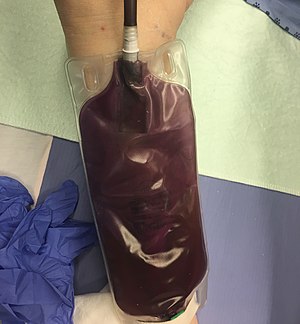
Purple urine in a catheterized patient with a UTI, https://upload.wikimedia.org/wikipedia/commons/thumb/4/49/PurpleUrine.jpg/300px-PurpleUrine.jpg
|
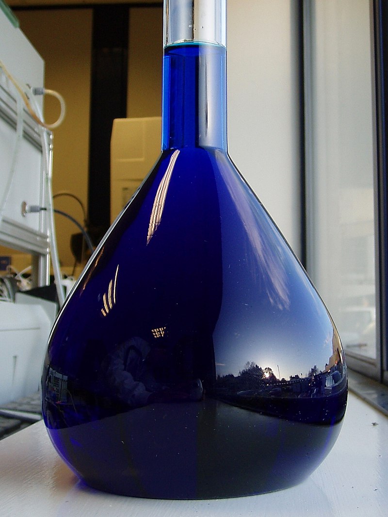
Methylene blue solution in a scan will turn the urine blue until it clears, https://upload.wikimedia.org/wikipedia/commons/thumb/a/a9/Reflections_in_a_flask_of_Methylene_Blue.jpg/800px-Reflections_in_a_flask_of_Methylene_Blue.jpg
|
Porphyria is a genetic disorder that turns the urine tea-colored or red, https://upload.wikimedia.org/wikipedia/commons/a/a8/Urine_of_patient_with_porphyria.png
|
Rhabdomyolysis, or muscle damage from trauma, M.S., or heart damage, can turn the urine tea or cola color, https://upload.wikimedia.org/wikipedia/commons/thumb/f/f7/RhabdoUrine.JPG/800px-RhabdoUrine.JPG
|
Taking phenazopyridine, or pyridiumurine, an over-the-counter anesthetic for bladder pain due to UTI, overactive bladder, spasms, or cystitis, will turn the urine bright orange, https://upload.wikimedia.org/wikipedia/commons/thumb/c/ca/Pyridiumurine.jpg/800px-Pyridiumurine.jpg
|
Bright yellow urine results from taking vitamin B or riboflavin supplements, https://upload.wikimedia.org/wikipedia/commons/thumb/a/a5/Micrococcus_riboflavin.jpg/1024px-Micrococcus_riboflavin.jpg
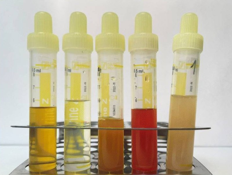
The normal color of urine is a pale straw color, which is a pale yellow, and normal turbidity should be clear. Urine that is darker yellow may signify dehydration, or the need to drink more water. If it is bright neon yellow, this signifies that the individual has taken a vitamin B supplement. If it is too pale, the individual is not concentrating the urine enough. This may or may not be significant. If it is a pattern, it can be significant. Urine that is orange contains bilirubin and sometimes urobilinogen. This could possibly signify liver issues, such as cirrhosis, hepatitis, toxicity, and/or jaundice. If it is cloudy, or turbid, something is in the urine that should not be there. If it is red, it may or may not signify an infection or presence of a kidney stone. If a female is menstruating, it can contaminate the urine with blood. Urine with pus is called pyuria, which is a combination of bacteria, cell debris, and white blood cells. This type of urinary tract infection typically smells very strong, often of ammonia. If the urine is blue, the person likely just had a scan or pyelogram where their veins were injected with blue dye. If the urine is green, they likely have a urinary tract infection with the organism Pseudomonas aeruginosa, a bacterium that produces the green pigment making the urine green. Sometimes consuming a large amount of asparagus can turn the urine green, and consuming a large amount of beets can make it appear red/pink or purple/red.
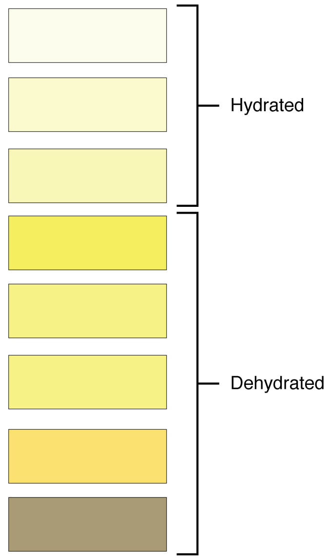
Urine colors
|
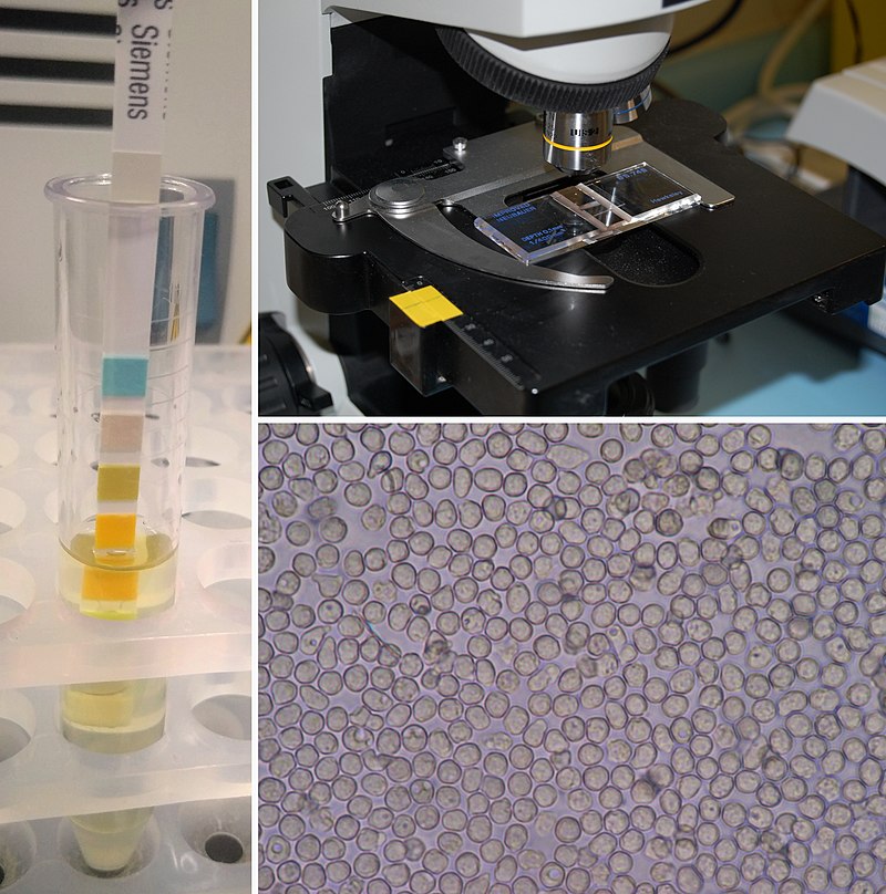
Urinalysis components
|
|
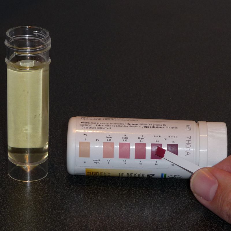
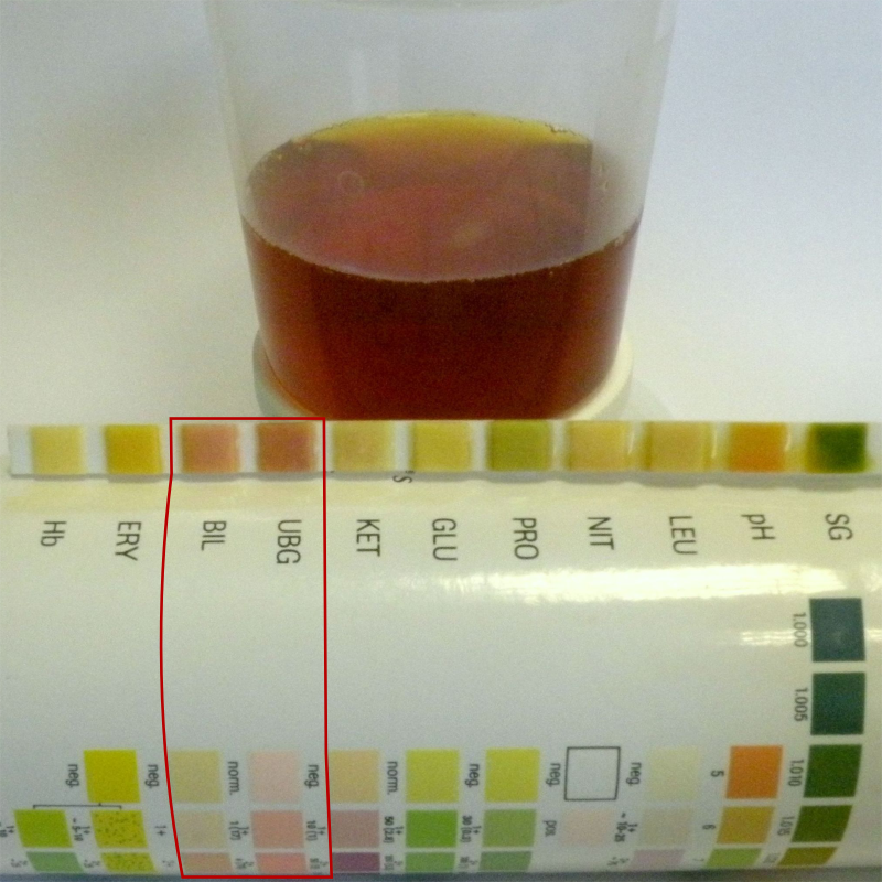
The ketones test strip is part of the urinalysis, but is also available as a stand-alone test. If ketones are present, this can either signify the ketone diet, or it can signify that a person with type 2 diabetes mellitus has glucose spilling over into the urine, making it acidic, and putting them closer to ketoacidosis, which can be a medical emergency.
|
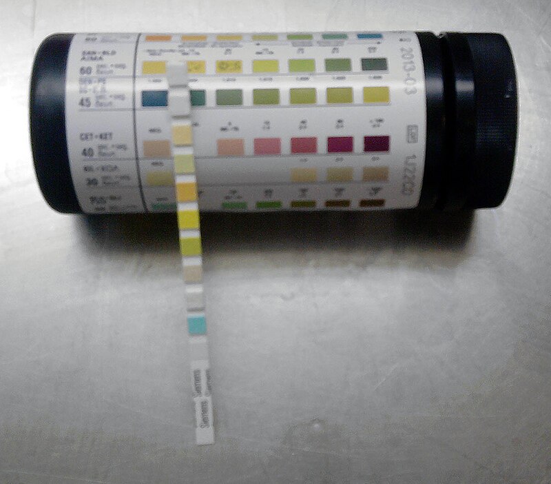
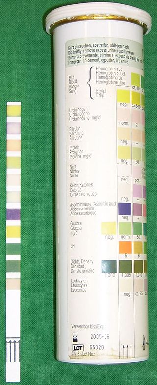
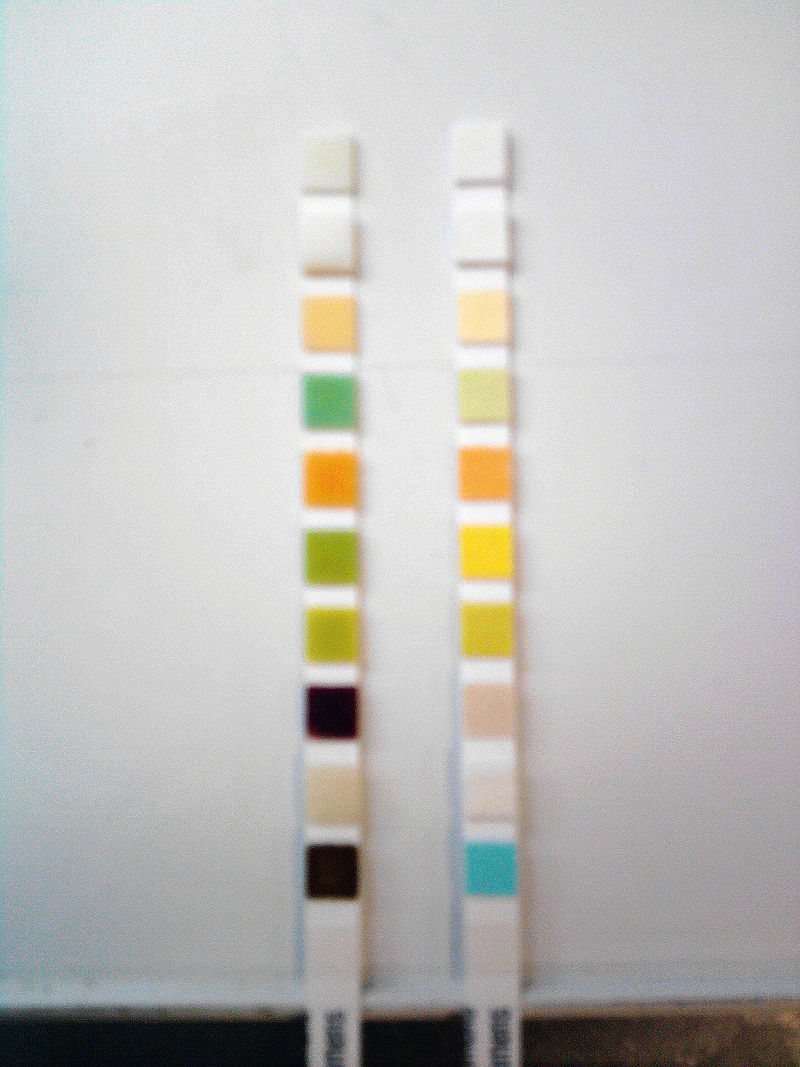
Here is an example of how to match up the multistix dip stick next to the color chart for interpretation.
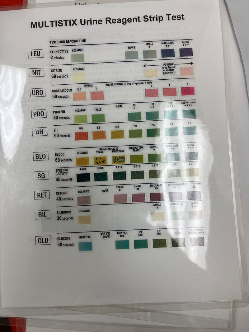
This guide shows the color-coded values to look for when interpreting the multistix urinalysis dip stix. It should be read from the bottom-up, starting with glucose and moving up, reading leukocytes last. The longer part with no pads is designed to serve as a handle to hold the strip. The canister contains this exact color chart for interpretation.
Urine dip stix are part of the urinalysis. They must be within date (not expired), protected from light, and protected from moisture, and each square should be read within its designated time frame.
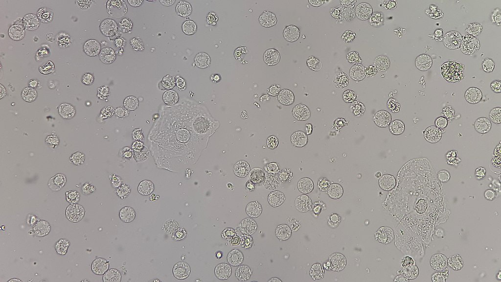
This urine contains many white blood cells, a couple of squamous epithelial cells, and some mixed bacteria. The number of white blood cells signifies inflammation, as well as possible infection, but the urinalysis strip test and culture will help decide that.
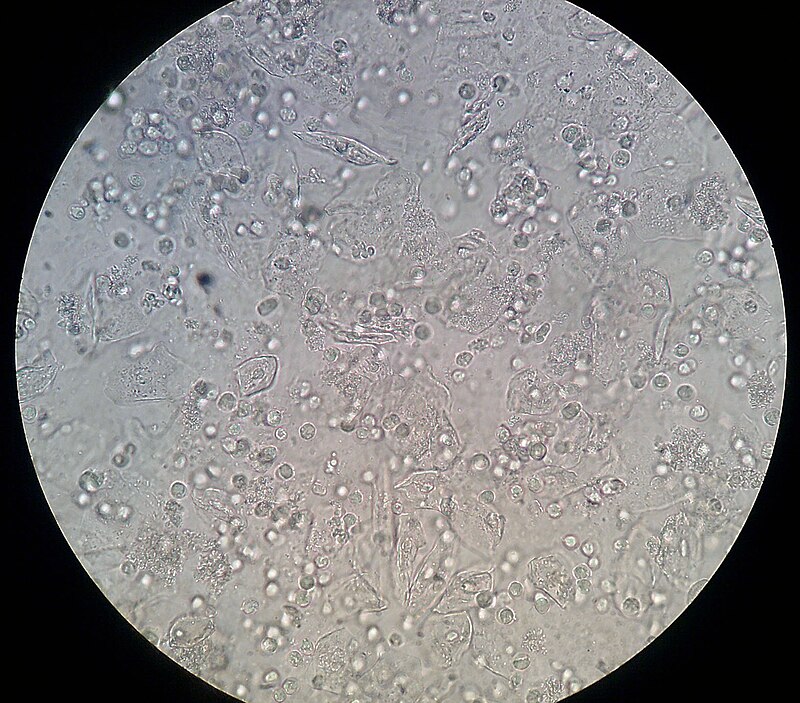
This is what squamous epithelial cells and white blood cells in the urine look like. This signifies some inflammation. If bacteria or yeast are present, it would signify a UTI.
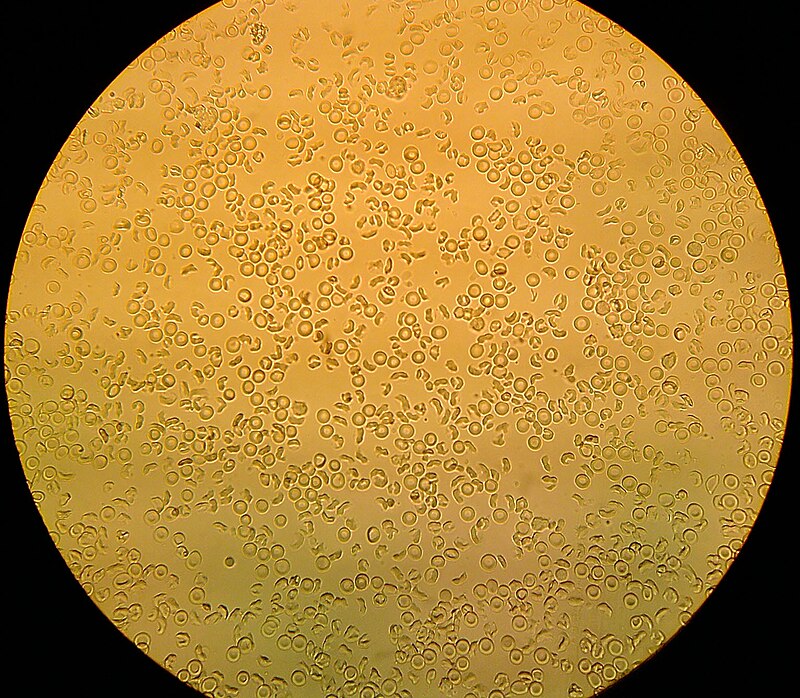
This is what red blood cells in the urine look like.
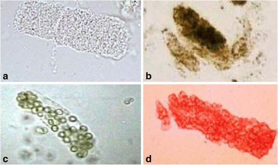
This slide shows 4 different types of casts in the urine. They form in the kidney tubules and signify either a kidney infection or a problem with the way that the kidneys are currently functioning.
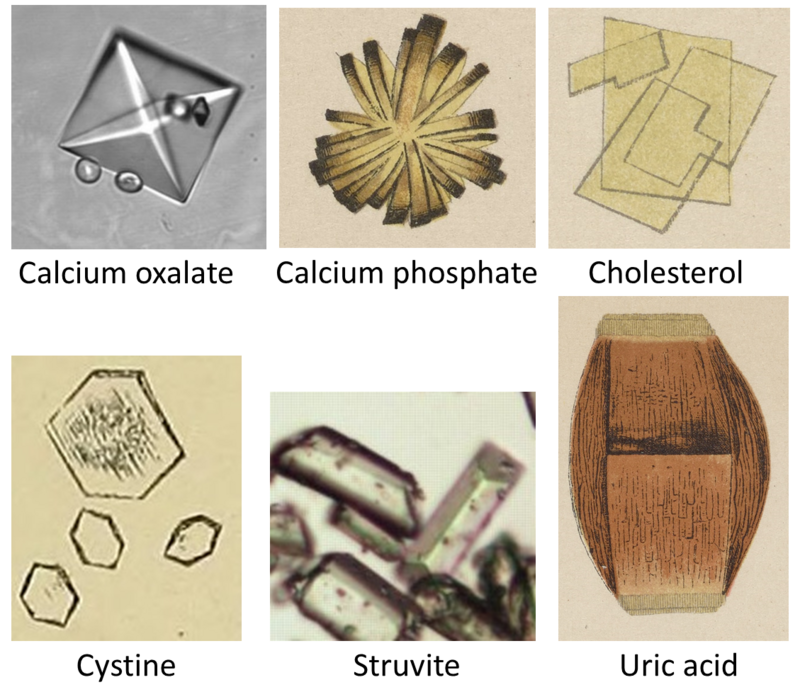
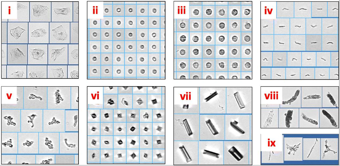
These are all different types of crystals that can be identified in urine. Some of them are normal, some are abnormal, and some can form when urine is exposed to air if it is not properly preserved within 2 hours of collection.
This IRIS cell urine analyzer is an automated machine with a built-in microscope, which can identify cells in the urine, bacteria, yeast, crystals, casts, and mucous.
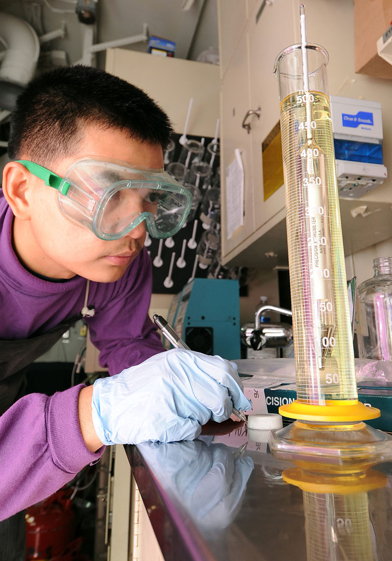
This Naval military student is learning how to read a urine hydrometer for specific gravity and is learning how to read a meniscus.
Pyruia is pus in the urine, consisting of white blood cells, cell debris, and bacteria. Here is the macroscopic collection, and you can see the microscopic values on the right. https://upload.wikimedia.org/wikipedia/commons/thumb/2/29/Pyuria2011.JPG/800px-Pyuria2011.JPG
|
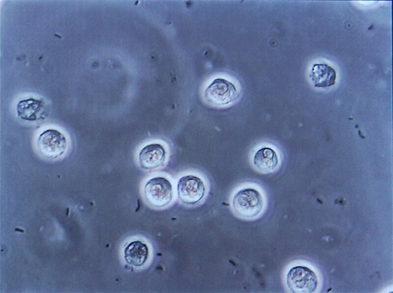
In this microscopic wet mount of concentrated urine sediment, you can see Gram-negative bacterial cells and lots of white blood cells (polymorphonuclear neutrophils), indicative of an infection.
|
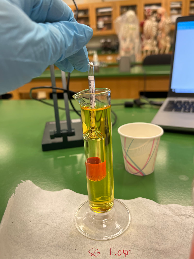
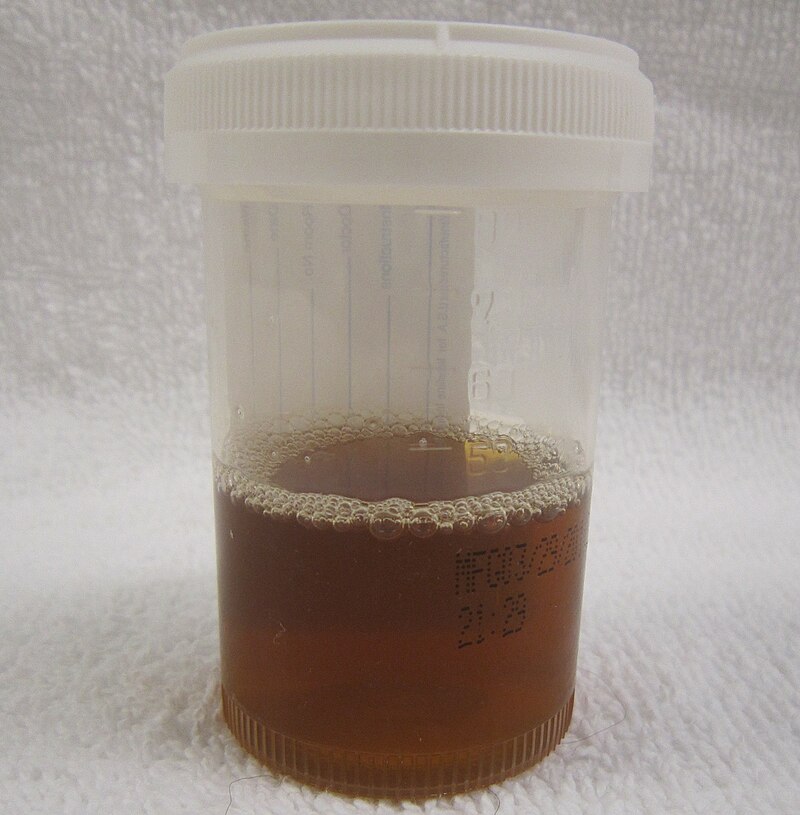
Author: Jeanette Reynolds, MS-Biology, M-ASCP, MLS-AMT, Certified in Emerging Diseases, Certified Instructor, Biology Instructor (Specific Gravity measured using a glass cylinder and glass hydrometer read at the meniscus); This one read 1.045, which is high. Normal SG should be between 1.001 and 1.030.
|
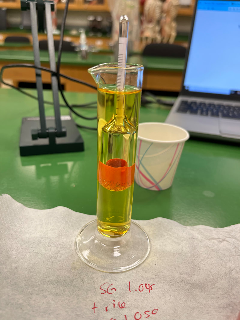
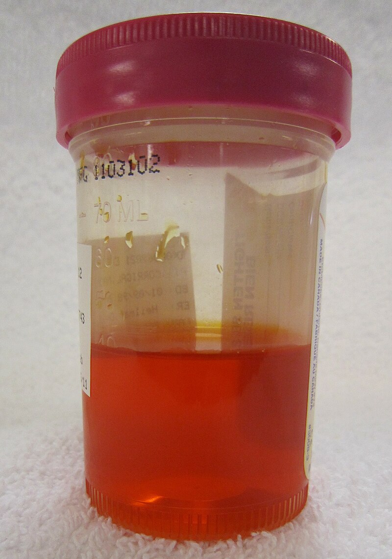
Author: Jeanette Reynolds, MS-Biology, M-ASCP, MLS-AMT, Certified in Emerging Diseases, Certified Instructor, Biology Instructor (Specific Gravity measured using a glass cylinder and glass hydrometer read at the meniscus); This one read 1.050, which is high. Normal SG should be between 1.001 and 1.030. NOTE: I added a rice grain to this one to represent glucose in the urine. It raised the SG from 1.045 to 1.050, which is very high. Glucose, proteins, lipids, cells, crystals, and/or casts, or kidney or bladder stones in the urine will raise the specific gravity.
|
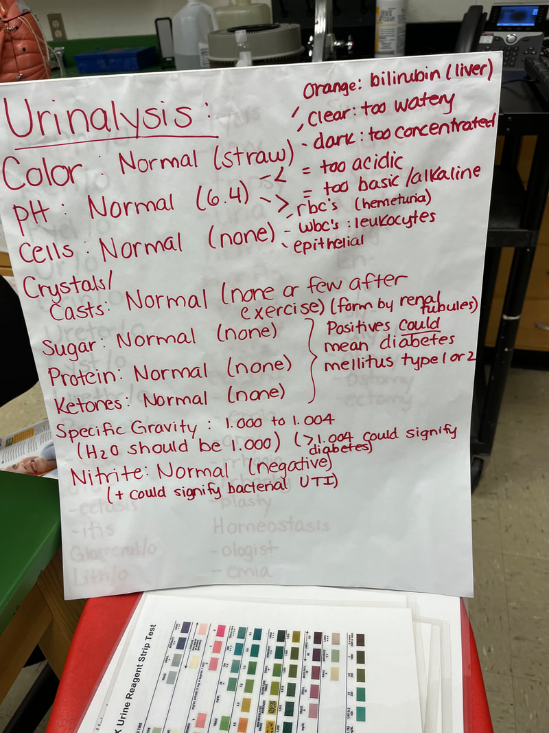
Author: Jeanette Reynolds, MS-Biology, M-ASCP, MLS-AMT, Certified in Emerging Diseases, Certified Instructor, Biology Instructor: Components of a urinalysis
|
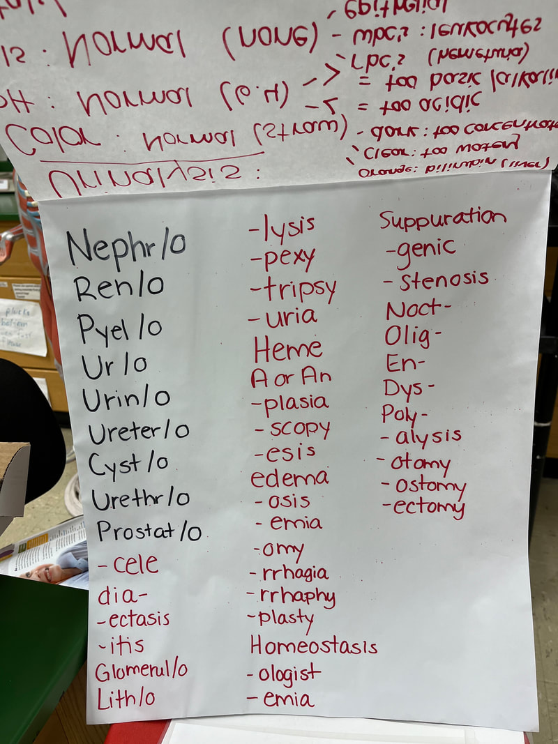
Author: Jeanette Reynolds, MS-Biology, M-ASCP, MLS-AMT, Certified in Emerging Diseases, Certified Instructor, Biology Instructor: Medical Terminology of the Urinary System
|
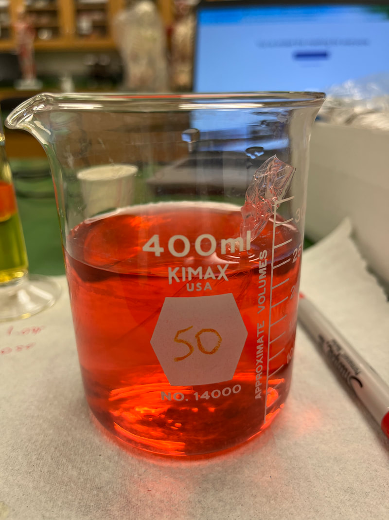
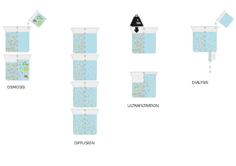
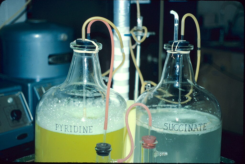
Author: Jeanette Reynolds, MS-Biology, M-ASCP, MLS-AMT, Certified in Emerging Diseases, Certified Instructor, Biology Instructor: Simulated kidney dialysis (The dialysis tubing has been tied off and filled with DI water to represent a cell, and the beaker is tap water containing minerals with negatively-charged dye. When solutes (solid molecules) cross through the porous dialysis tubing, representing the porous selectively permeable cell membrane, this is known as diffusion. In simple diffusion, such as the drop of dye evenly dispersing into the water, no help is needed, no energy is needed, and the solutes move down the concentration gradient. Those that need a little help of a channel or carrier protein are known as facilitated diffusion. Osmosis is when water moves across the cell membrane to balance out hypertonic or hypotonic solutions to cells so that they don't crenate (shrink) or swell and possibly lyse (burst open).
|
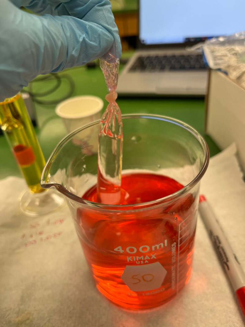
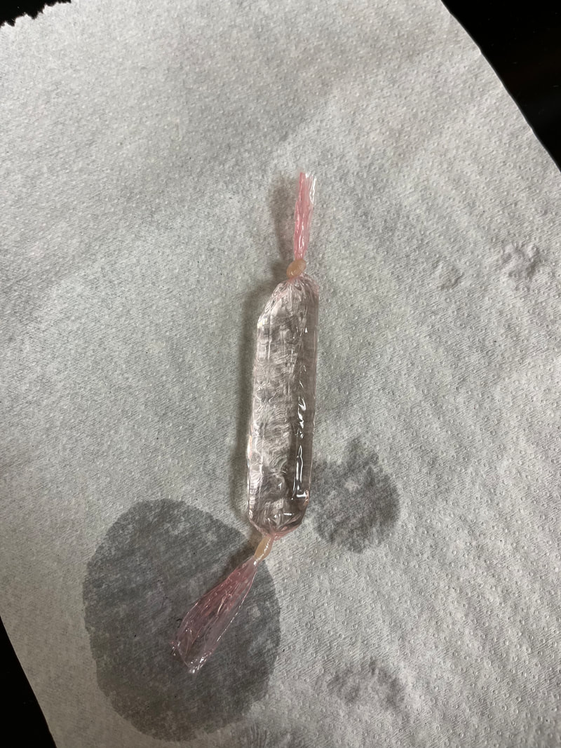
Author: Jeanette Reynolds, MS-Biology, M-ASCP, MLS-AMT, Certified in Emerging Diseases, Certified Instructor, Biology Instructor: Simulated kidney dialysis (The dialysis tubing has been tied off and filled with DI water to represent a cell, and the beaker is tap water containing minerals with negatively-charged dye. When solutes (solid molecules) cross through the porous dialysis tubing, representing the porous selectively permeable cell membrane, this is known as diffusion. In simple diffusion, such as the drop of dye evenly dispersing into the water, no help is needed, no energy is needed, and the solutes move down the concentration gradient. Those that need a little help of a channel or carrier protein are known as facilitated diffusion. Osmosis is when water moves across the cell membrane to balance out hypertonic or hypotonic solutions to cells so that they don't crenate (shrink) or swell and possibly lyse (burst open). In this case, you can see after 30 minutes that the dye crossed the cell membrane and went into the cell. This is because the negatively-charged dye molecules are attracted to the positive charges of the H+ ions in the water, so some dye molecules moved across the cell membrane to bond to the H+ ions found within water.
|
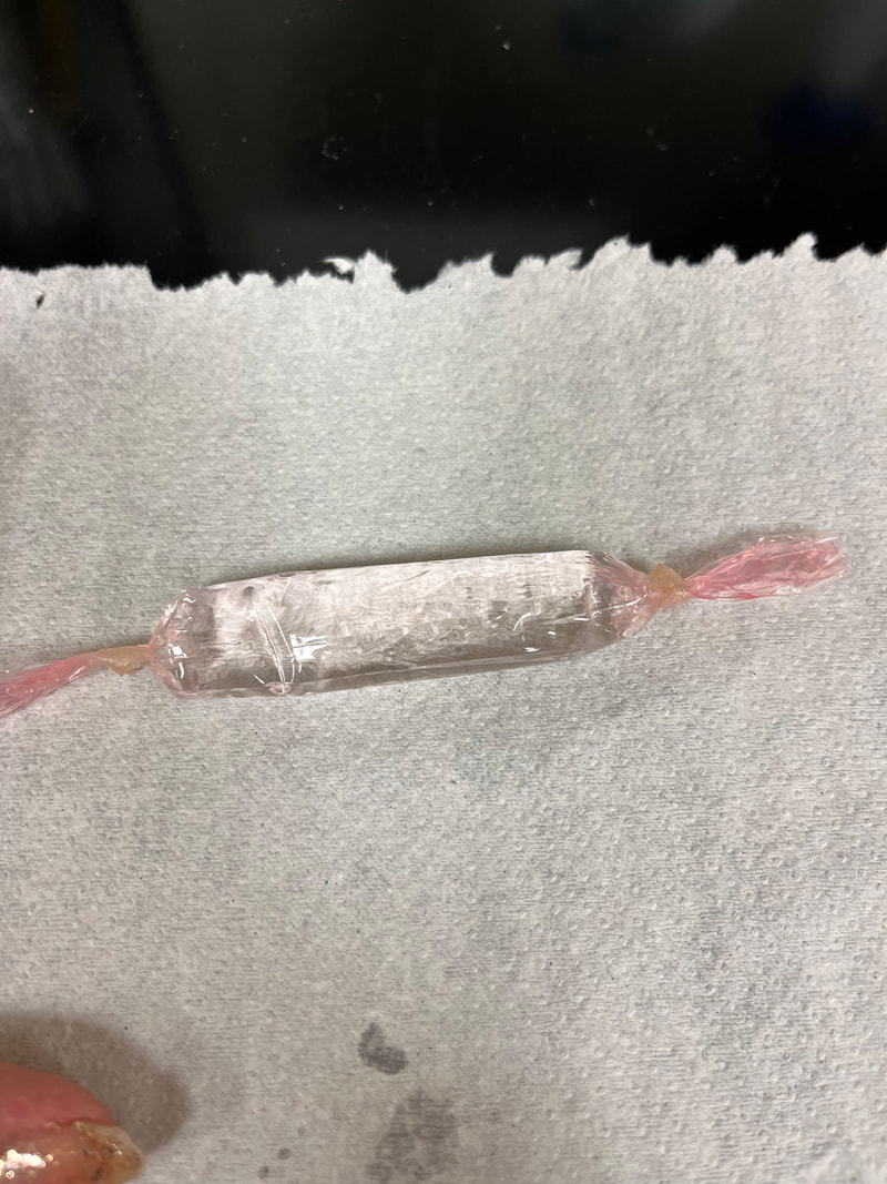
Author: Jeanette Reynolds, MS-Biology, M-ASCP, MLS-AMT, Certified in Emerging Diseases, Certified Instructor, Biology Instructor: Simulated kidney dialysis (The dialysis tubing has been tied off and filled with DI water to represent a cell, and the beaker is tap water containing minerals with negatively-charged dye. When solutes (solid molecules) cross through the porous dialysis tubing, representing the porous selectively permeable cell membrane, this is known as diffusion. In simple diffusion, such as the drop of dye evenly dispersing into the water, no help is needed, no energy is needed, and the solutes move down the concentration gradient. Those that need a little help of a channel or carrier protein are known as facilitated diffusion. Osmosis is when water moves across the cell membrane to balance out hypertonic or hypotonic solutions to cells so that they don't crenate (shrink) or swell and possibly lyse (burst open). In this case, you can see after 30 minutes that the dye crossed the cell membrane and went into the cell. This is because the negatively-charged dye molecules are attracted to the positive charges of the H+ ions in the water, so some dye molecules moved across the cell membrane to bond to the H+ ions found within water, turning the cell slightly pink. This is an example of either simple or facilitated diffusion.
|

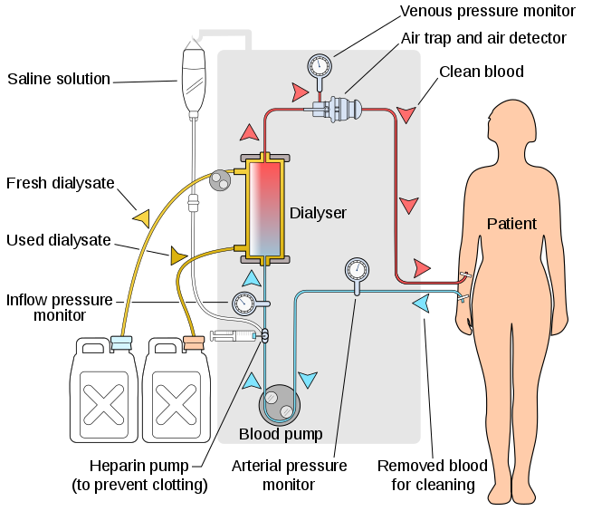
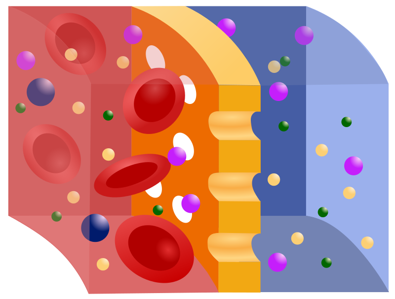
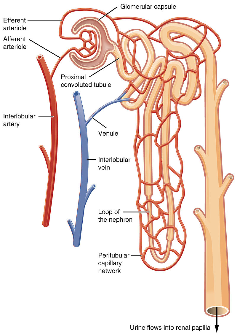
This is a great example of the structure of the human kidney. The filtration units of the kidneys are known as the nephrons. They make the urine and filter it. Each nephron is wrapped with special capillaries one-cell thick called glomeruli, which enable substances to cross in and out of the membrane. When diabetic individuals have protein or glucose spill over into the urine, it is because the large molecules have stretched out the glomeruli of the nephrons of the kidneys, resulting in a spilling over into the urine, which is not normal. This can damage the kidneys over time, resulting in the need to rely upon dialysis to do the work of the kidneys.
|
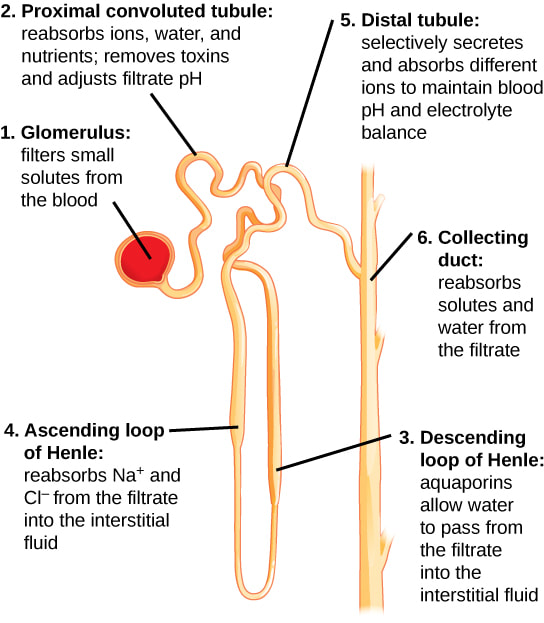
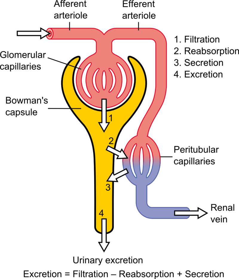
Urine is made in the nephrons of the kidney, represented by #1 here. You can see the route it follows, as water is reabsorbed, toxins are removed, the pH is adjusted, and ions are also reabsorbed. This occurs throughout the tubules of the kidneys.
|
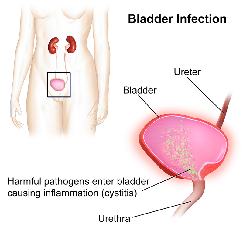
https://upload.wikimedia.org/wikipedia/commons/thumb/e/e5/Bladder_Infection.png/800px-Bladder_Infection.png
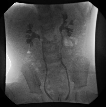
Urinary pyelogram showing reflux from the ureters back into the renal pelvices.https://upload.wikimedia.org/wikipedia/commons/0/0a/Vesicoureteral-reflux-004.jpg
Fluid and Electrolyte Balance:
Lecture Objectives, Chapter 19: Fluid and Electrolyte Balance
Upon completion of this chapter, you should be able to:
Upon completion of this chapter, you should be able to:
- List, describe and compare and contrast the body fluid compartments and their subdivisions.
- Discuss avenues by which water enters and leaves the body and the forces that move fluids into and out of the blood.
- Explain the mechanisms used by the body to maintain fluid balance.
- Discuss the nature and importance of electrolytes in body fluids.
- Describe examples of common fluid and electrolyte imbalances.
- Define the terms fluid balance, electrolytes, ions, electrolyte balance, hypernatremia, hyponatremia, edema, hyperkalemia, hypokalemia, hypercalcemia, hypocalcemia, diuretic, dehydration, overhydration, water intoxication, cations, anions, intracellular fluid, interstitial fluid, extracellular fluid.
Urinalysis:
Urinalysis - an analysis of the physical, chemical, and microscopic characteristics of urine and a measure of urine volume. See Table 28.1 for normal characteristics of urine.
Urine Volume - varies depending on the water content of the body, and decreases when body fluid volume is low.
Specific Gravity of Urine - the weight of a volume of urine divided by the weight of the same volume of distilled water.
- Urine weight per volume is higher than distilled water because of the presence of solutes in urine.
- The more solutes present in urine (protein, sugar, etc...), the higher the specific gravity.
- Normal urine: 95% water and 5% solutes, including electrolytes, urea, creatinine, uric acid, and metabolic end products of hormones and other substances.
- Electrolytes: sodium, potassium chloride, magnesium, and other ions
- Urea: formed from breakdown of amino acids from proteins
- Creatinine: formed from breakdown of creatine phosphate in muscle tissue
- Uric acid: formed from breakdown of nucleic acids
- Electrolytes: sodium, potassium chloride, magnesium, and other ions
Acid-Base Balance:
Lecture Objectives, Chapter 20, Acid-Base Balance:
- Define the term "acid-base balance" and discuss the concept of pH.
- Define the terms acid, base, buffer, buffer pair, pH, acidosis, alkalosis.
- Compare and contrast strong and weak acids and bases.
- Describe the role of a buffer.
- Contrast the respiratory and urinary mechanisms of pH control.
- Compare and contrast metabolic and respiratory types of alkalosis and acidosis and pH imbalances.
- Discuss compensatory mechanisms that may help return blood pH to near-normal values in cases of pH imbalances.
Lab Objectives, Acidosis and Alkalosis:
Upon completion of this exercise, you should be able to:
Alkalosis and Acidosis Research Lab:Conditions:
Respiratory Acidosis
Respiratory Alkalosis:
Metabolic Acidosis and Diabetic Ketoacidosis:
Metabolic Alkalosis:
Causes:
Signs and Symptoms:
Prevention and Treatment:
pH:
Upon completion of this exercise, you should be able to:
- Differentiate between respiratory alkalosis and acidosis, their causes, and signs and symptoms.
- Differentiate between metabolic alkalosis, metabolic acidosis, and diabetic ketoacidosis, recognize the signs and symptoms, and identify the causes.
- Understand the role of pH, acids, bases, and buffers in the human body.
Alkalosis and Acidosis Research Lab:Conditions:
Respiratory Acidosis
Respiratory Alkalosis:
Metabolic Acidosis and Diabetic Ketoacidosis:
Metabolic Alkalosis:
Causes:
Signs and Symptoms:
Prevention and Treatment:
pH:
