Nerves:
Lecture Objectives: Chapter 9Upon completion of the lectures and chapters, you should be able to:
Upon completion of the lab exercises, you should be able to:
- List the organs and divisions of the nervous system, and describe the generalized functions of the system as a whole.
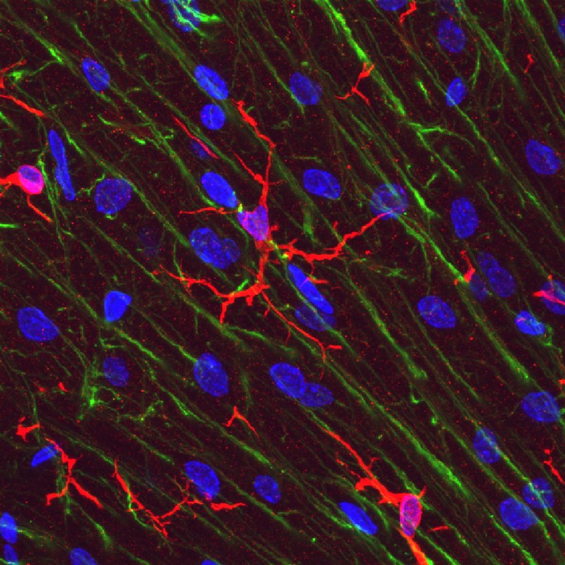
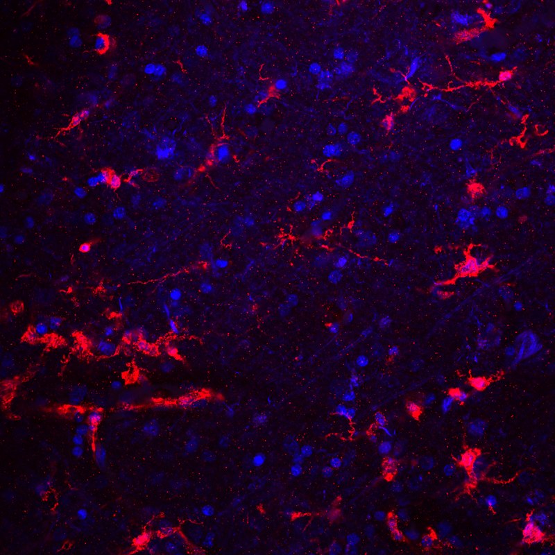
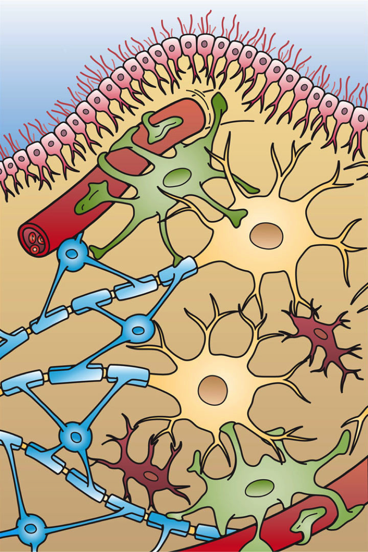
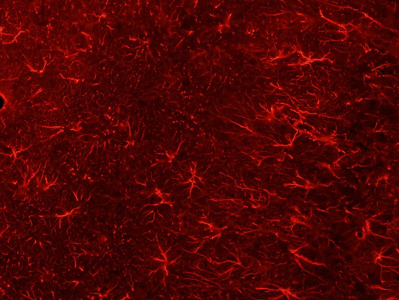
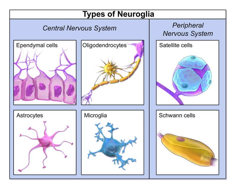
- Identify the major types of cells in the nervous system and discuss the functions of each.
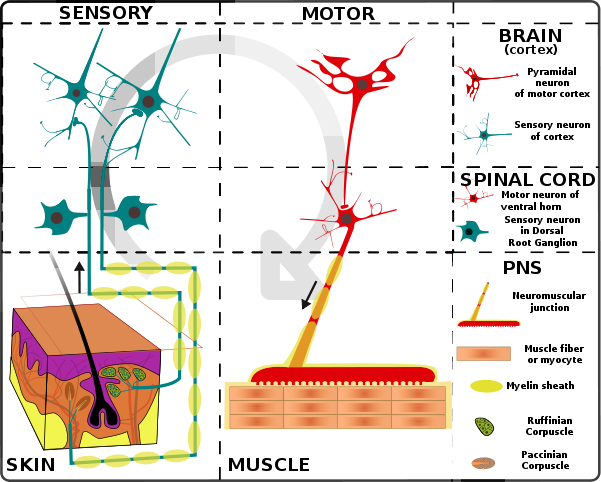
- Identify the anatomical components of a reflex arc and explain its function.
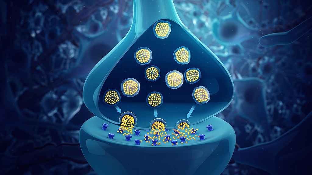
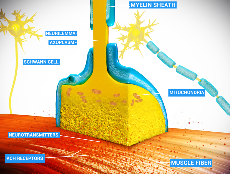
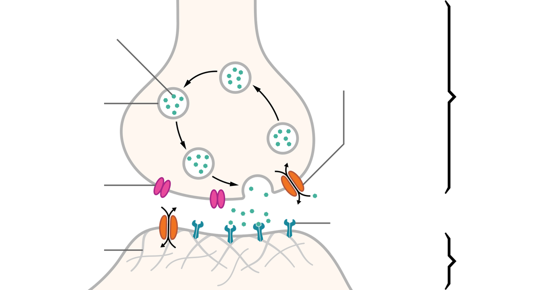
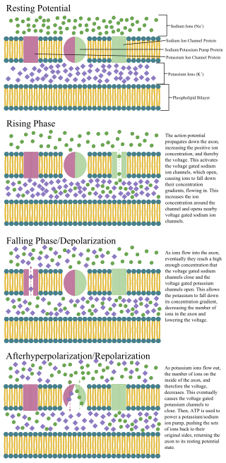
- Explain the mechanisms of transmission of a nerve impulse along a nerve fiber and across a synapse.
- Identify the major anatomical components of the brain and spinal cord, and briefly comment on the functions of each.
- Compare and contrast cranial and spinal nerves.
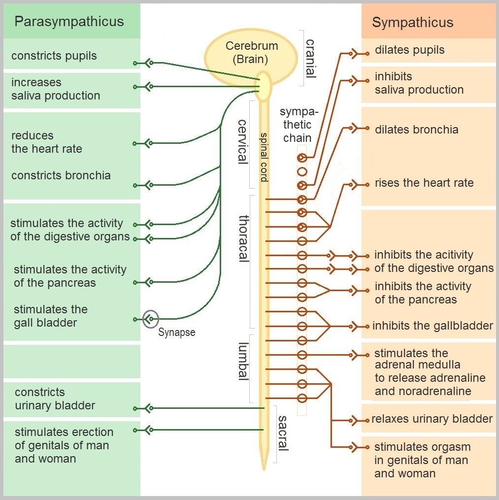
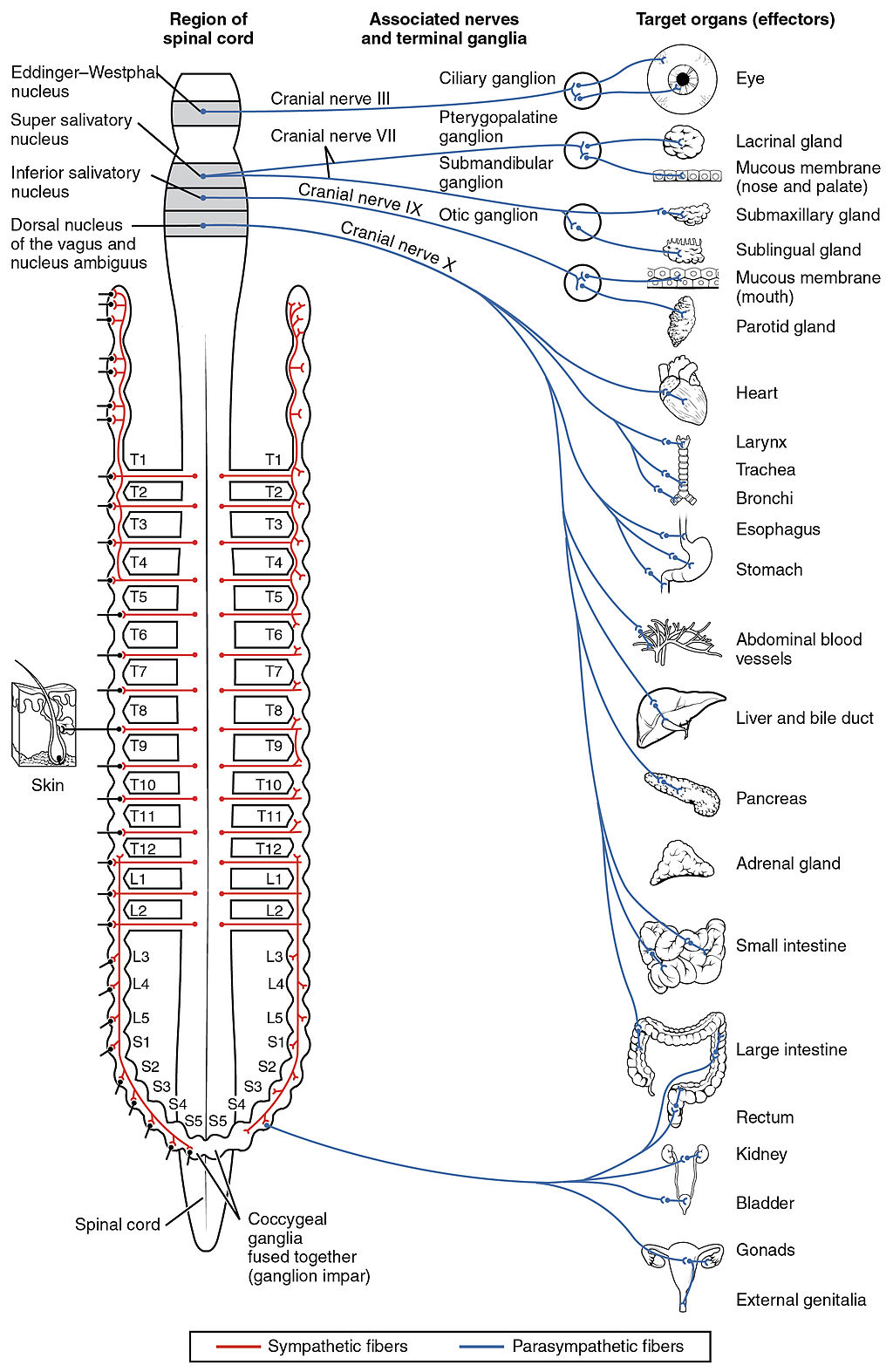
- Discuss the anatomical and functional characteristics of the two divisions of the autonomic nervous system.
- Classify the sense organs as general or special and explain the basic differences between the two groups.
- Discuss how a stimulus is converted into a sensation.
Upon completion of the lab exercises, you should be able to:
- Identify the external and internal features of the spinal cord on models or charts.
- Name the four spinal divisions of a spinal cord.
- Name the spinal plexuses and the major nerves arising from each plexus, and identify them on a model or chart.
- Identify and describe the five components of a somatic reflex arc on models or charts.
- Explain the significance of clinical testing of reflexes.
- Identify the major structures of the brain on models and charts and describe their functions.
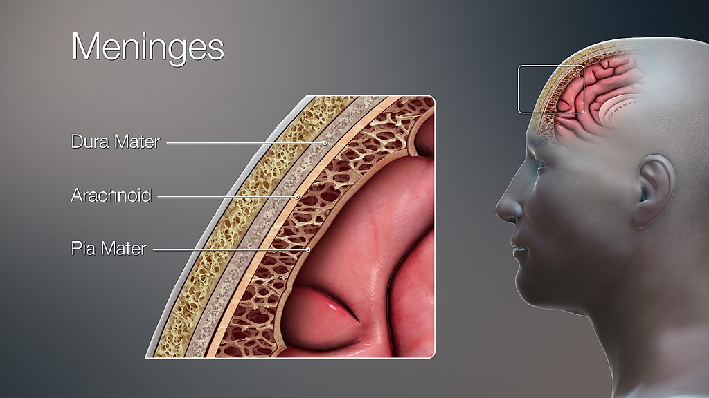
- Name the three meninges and describe their similarities and differences.
- Explain the production of cerebrospinal fluid and trace its circulation.
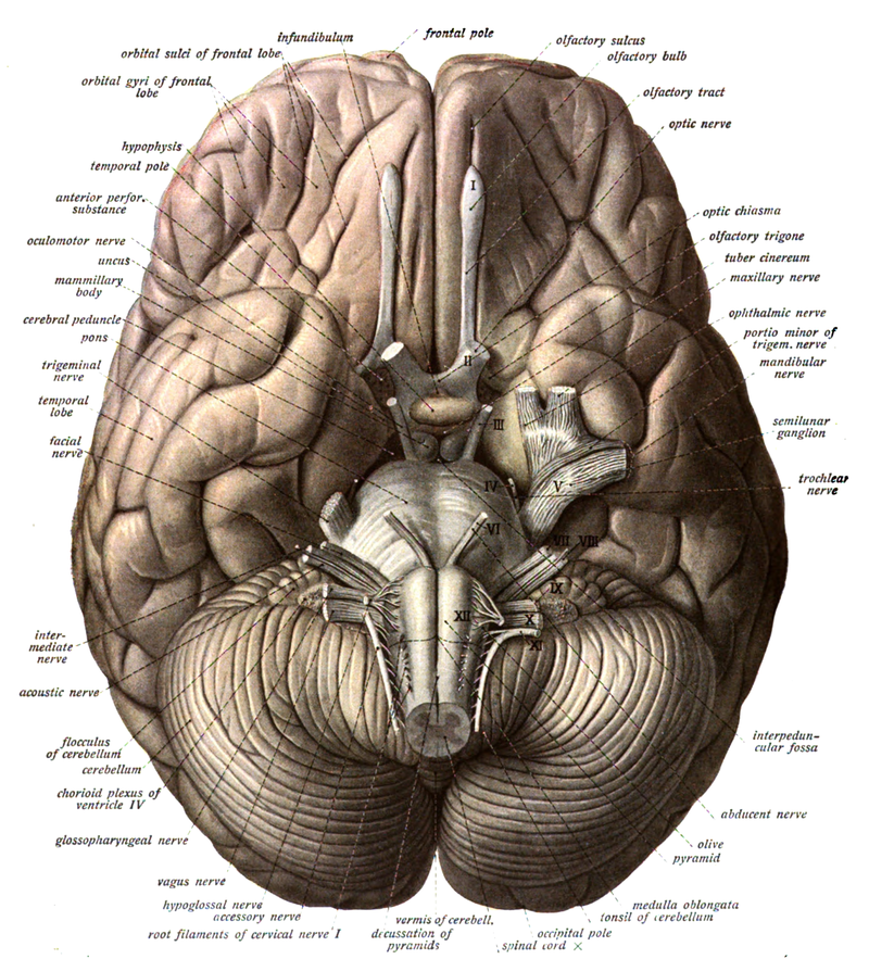
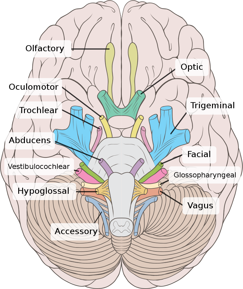
- Identify the 12 pairs of cranial nerves by name and Roman numeral on brain models and illustrations.
- State the function of the 12 pairs of cranial nerves.
- Compare the anatomy of the sheep brain with that of the human brain.
- Dissect the sheep brain and identify the lobes of the brain, the dura mater, the types of matter, the brain regions, the sulca and gyra, and the fissures.
Brain and Spinal Meninges:
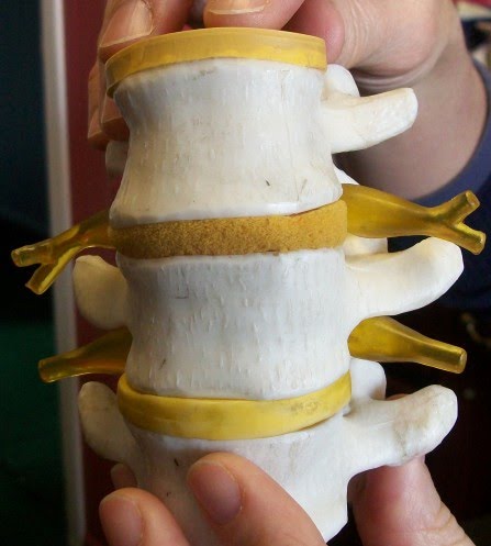
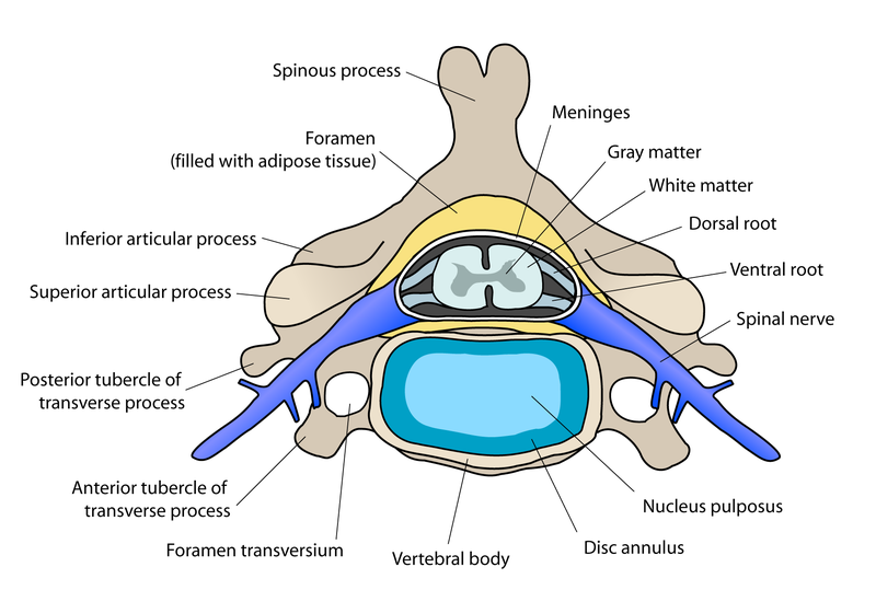
Adipose Tissue: padding of adipocytes (lipids) that cushion the spinal cord in the epidural space
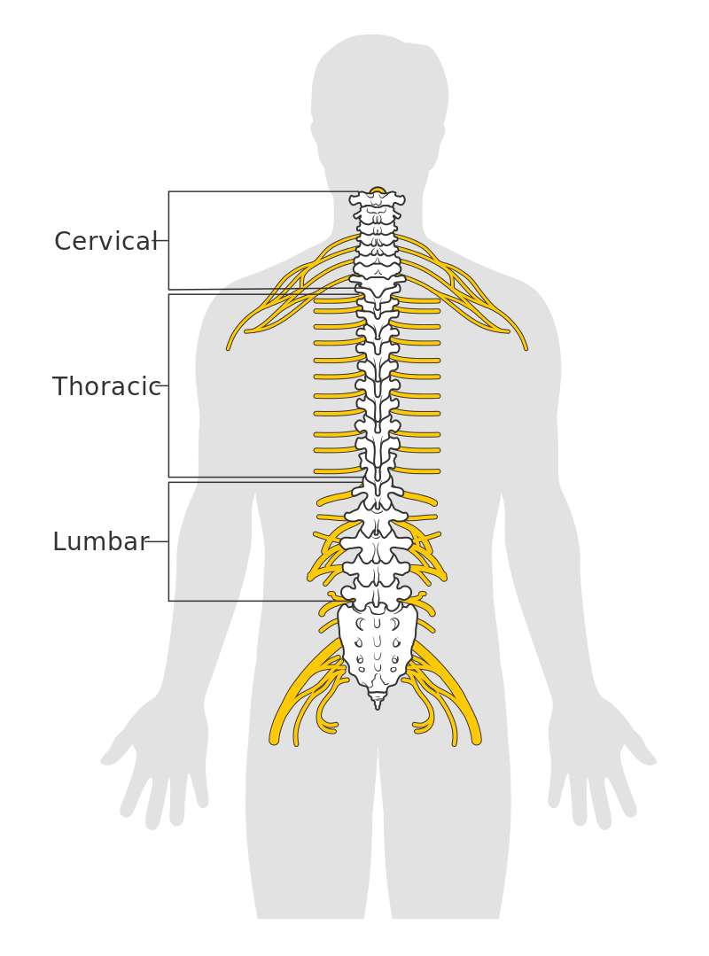
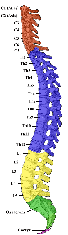
Spinal Vertebrae:
31 pairs total
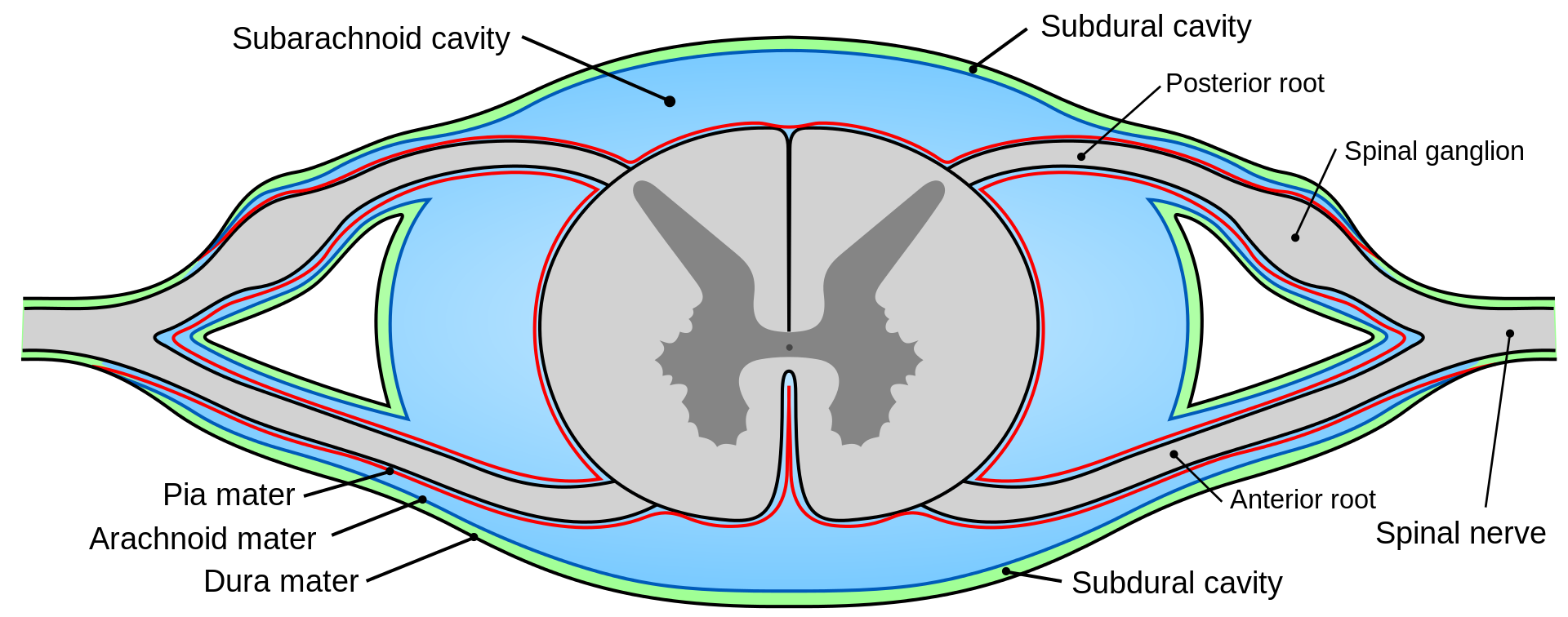
- Pia Mater: inner meninx; delicate; hugs the spinal cord
- Dura Mater: tough, outer meninx; single-layered; deep to the adipose tissue in the epidural space; superficial to the spiderweb-like arachnoid mater
- Arachnoid Mater: spiderweb-like layer in the middle, which makes the CSF
Adipose Tissue: padding of adipocytes (lipids) that cushion the spinal cord in the epidural space
Spinal Vertebrae:
- Intervertebral foramen/foramina: spaces in the spinal vertebrae through which spinal nerves emerge and exit
- Foramen magnum: large foramen space in the base of the skull (occipital) where the spinal cord begins
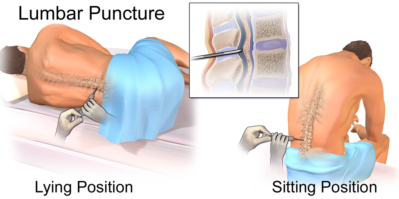
- L1-L2: spinal cord ends between these 2 vertebrae
- Begins at the foramen magnum
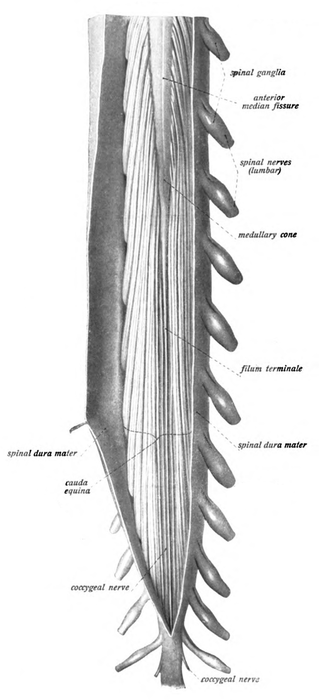
- Conus medullaris: ends inferiorly at L1-L2 as this structure
- Filum terminale: extension of the pia mater that continues past the conus medullaris, which connects the spinal cord to the coccyx
- Cauda equina: nerves arising from the inferior portion of the spinal cord that continue ("tail" or "horse's tail"); this is the tapered end of the spinal cord
- Plexus: distal to where a spinal nerve passes through its intervertebral foramen; anterior branches form a braided network prior to innervating the body structures; There are 4: cervical, brachial, lumbar, sacral (NOTE: thoracic nerves T2-T12 DO NOT form plexuses)
31 pairs total
- 8 pairs cervical
- 12 pairs thoracic
- 5 pairs lumbar
- 5 pairs sacral
- 1 coccygeal
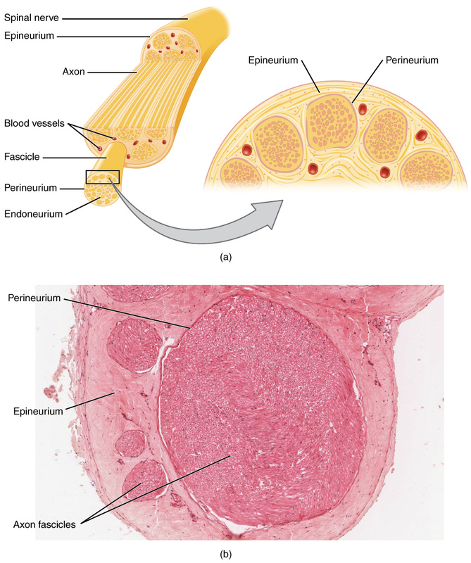
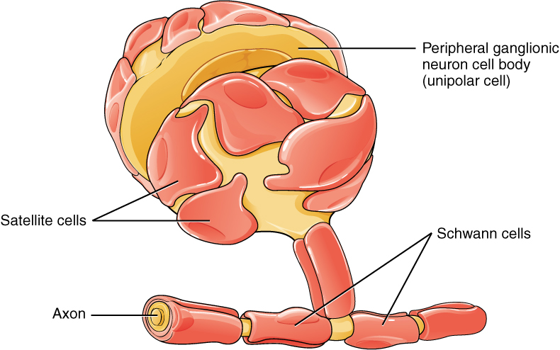
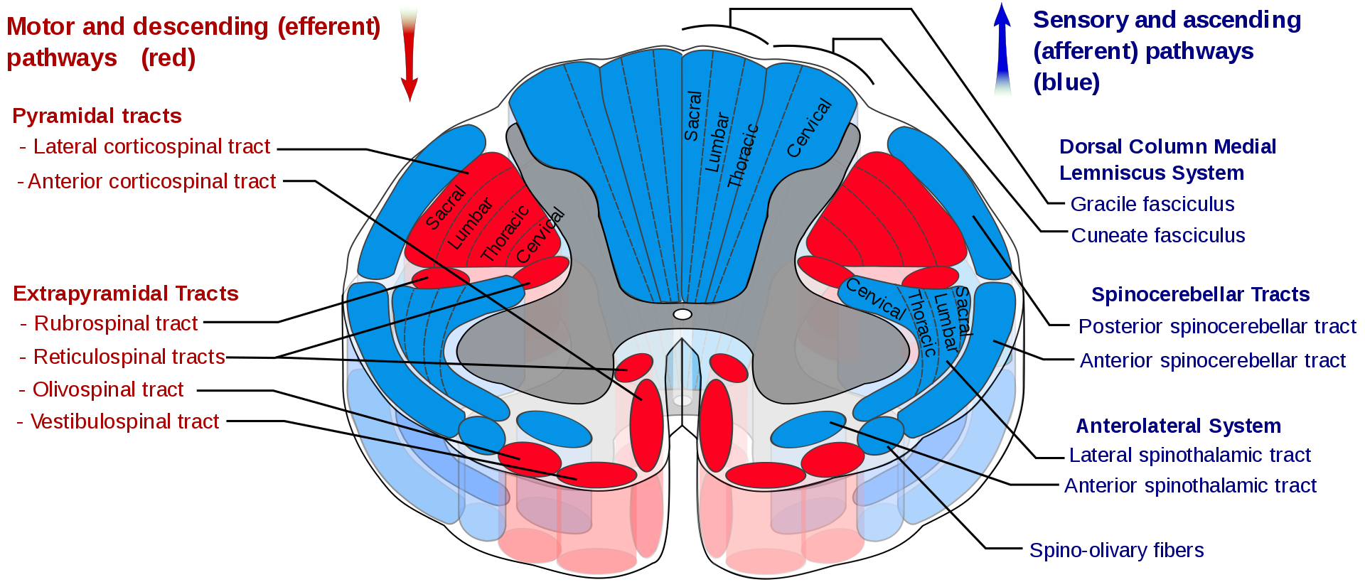
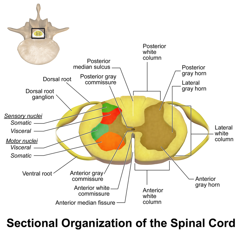
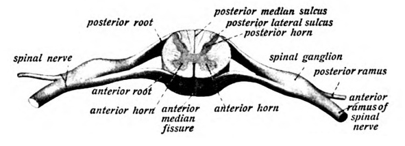
Gray Matter: contains neuron cell bodies and unmyelinated processes
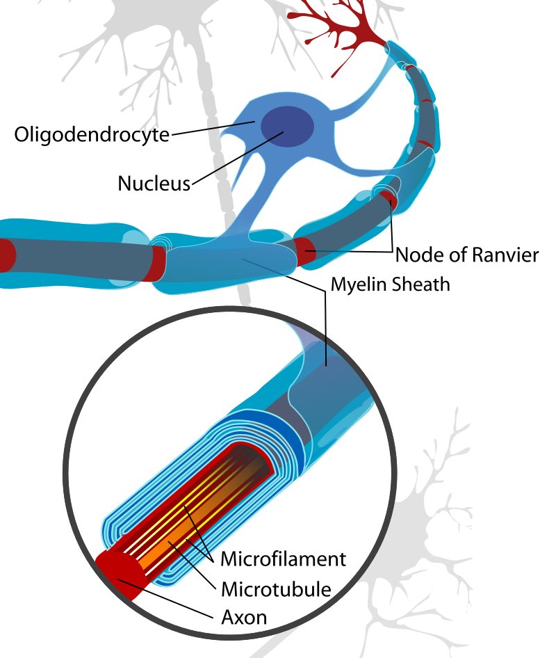
White Matter: contains myelinated axons (myelin sheath composed of Schwann cells and Nodes of Ranvier)
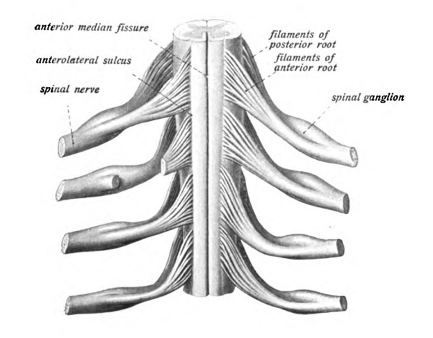
Anterior (ventral) root: sensory branch entering the spinal cord
Posterior (dorsal) root: motor branch exiting the spinal cord
Posterior root ganglion: swelling that contains sensory neuron cell bodies; some viruses can survive in a dormant state here
Central canal: small round passageway of the spinal cord containing the CSF
Filum terminale: extension of the pia mater that attaches the spinal cord to the coccyx
Conus medullaris: end of spinal cord between L1-L2
Reflexes and Reflex Arcs: Somatic:
Reflex: rapid, involuntary motor response to a stimulus (hot, cold, pain, pressure, tickle, itch); response of the effector to stimulation by the motor neuron of the reflex arc
Reflex arc: neural pathway TO an effector; 5 components (in order) are as follows:
Autonomic visceral reflex: motor response involves cardiac or smooth muscle tissue or glands
Spinal reflex: mediated by spinal nerves
Cranial reflexes: mediated by cranial nerves
White Matter: contains myelinated axons (myelin sheath composed of Schwann cells and Nodes of Ranvier)
Anterior (ventral) root: sensory branch entering the spinal cord
Posterior (dorsal) root: motor branch exiting the spinal cord
Posterior root ganglion: swelling that contains sensory neuron cell bodies; some viruses can survive in a dormant state here
Central canal: small round passageway of the spinal cord containing the CSF
Filum terminale: extension of the pia mater that attaches the spinal cord to the coccyx
Conus medullaris: end of spinal cord between L1-L2
Reflexes and Reflex Arcs: Somatic:
Reflex: rapid, involuntary motor response to a stimulus (hot, cold, pain, pressure, tickle, itch); response of the effector to stimulation by the motor neuron of the reflex arc
Reflex arc: neural pathway TO an effector; 5 components (in order) are as follows:
- Sensory receptor (if stimulus is strong enough, it triggers an action potential in the sensory neuron)
- Sensory neuron (propagates the action potential and synapses with neurons in the spinal cord or brain)
- Integrating center (located within the gray matter of the CNS; transfers information from the sensory neuron to the motor neuron)
- Motor neuron (carries the action potential inititated by the integrating center to the effector)
- Effector (can be skeletal muscle, cardiac muscle, smooth muscle, or glands)
Autonomic visceral reflex: motor response involves cardiac or smooth muscle tissue or glands
Spinal reflex: mediated by spinal nerves
Cranial reflexes: mediated by cranial nerves
Patellar Reflex:
Patellar reflex: extension of the knee that occurs when the patellar tendon is stretched
Patellar reflex arc: sensory receptors in quadriceps femoris muscle group; tap it to stimulate muscle spindles to initiate nerve impulses in axons of sensory neurons
Sensory neuron: sensory axons carry nerve impulses to integration center (gray matter) in spinal cord
Integrating center: sensory axons carry nerve impulses to the IC in gray matter in spinal cord, where they synapse with and initiate the nerve impulses in the motor neuron innervating the quadriceps muscle gropu
Motor neuron: axons travel in the femoral nerve to the quadriceps
Effector: quadriceps contracts and extends the leg when stimulated
Clasping Hands and Pulling Hands: reinforces/enhances the patellar reflex as the patellar tendon is stimulated
Clenching the Teeth: reinforces/enhances the biceps reflex
Biceps reflex:
Biceps reflex: contraction of biceps brachii muscle when the biceps tendon is stretched; stimulates tension in the biceps brachii.
Deep Tendon Reflex: The calcaneal tendon is stimulated and contracts during this test.
Plantar Flexion: Babinski Reflex
Biceps reflex: contraction of biceps brachii muscle when the biceps tendon is stretched; stimulates tension in the biceps brachii.
Deep Tendon Reflex: The calcaneal tendon is stimulated and contracts during this test.
Plantar Flexion: Babinski Reflex
In this test, cutaneous receptors are stimulated. Since nerves are still developing in infants, the babinski sign is normal. In adults, however, with mature nerves, the babinski sign is pathological and indicates neurological damage.
THE BRAIN:
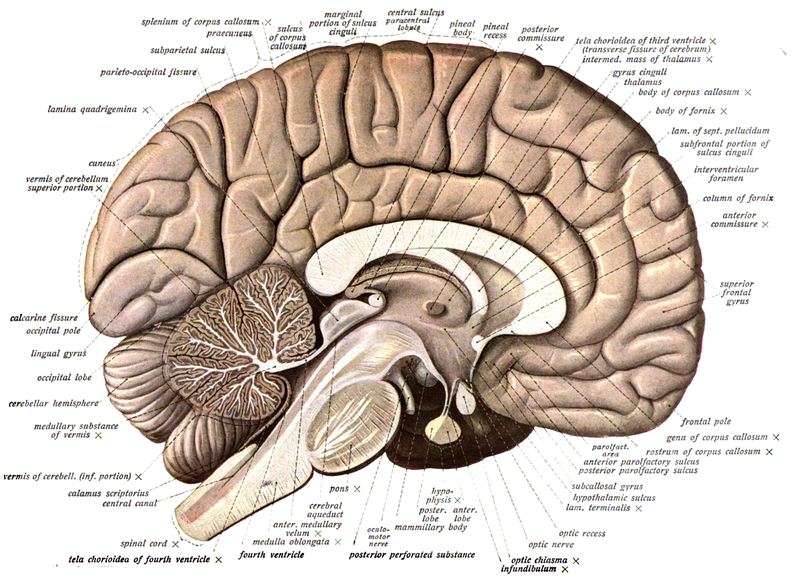
Major Brain Regions, Midsagittal View:
Major Brain Regions:
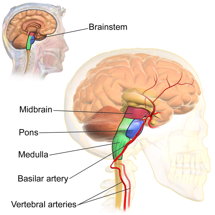
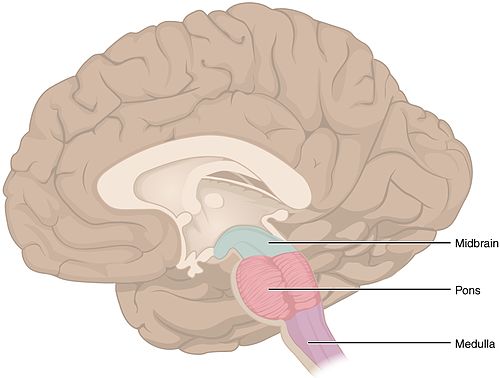
1. Brain Stem: connected to the superior part of the spinal cord and the diencephalon
3 Parts: Midbrain, Pons, Medulla Oblongata
4. Cerebrum: largest portion; envelops the diencephalon; dominant structure; made up of right and left hemispheres connected by the corpus callosum; largest and most complex division of the brain; superior to and surrounding the diencephalon and part of the brain stem; center of higher mental processes and learning, intelligence, communication, memory, reasoning, emotions, interpretation of sensory input, initiation of skeletal muscle contraction; 4 lobes:
External Anatomical Features:
White Matter: lies deep to the outer cortex and is made mostly of myelinated axons
Corpus Callosum: observable in midsagittal sections of the brain; a band of myelinated axons that connects the 2 cerebral hemispheres
THE BRAIN:
Major Brain Regions, Midsagittal View:
Major Brain Regions:
1. Brain Stem: connected to the superior part of the spinal cord and the diencephalon
3 Parts: Midbrain, Pons, Medulla Oblongata
- Midbrain: smaller area superior to the pons and inferior to the diencephalon; contains nerve tracts that connect the upper and lower brain areas
- Pons: expanded swelling located superior to the medulla oblongata and anterior to the cerebellum; respiratory centers that aid the medulla oblongata in controlling breathing/respiration; relays information to the diencephalon and cerebellum
- Medulla oblongata: superior to the spinal cord; most vital part of the brain that keeps us alive; respiratory and cardiovascular control centers that control rate and depth of breathing, rate and force of heartbeat and blood pressure relfexes; contains sensory and motor tracts that relay information between spinal cord and higher brain centers
- Thalamus: inner chamber; egg-shaped bodies, paired; take up 80% of the diencephalon; Grand Central relay station for sensory fibers that synapse there to relay information to particular areas of the cerebral cortex to be interpreted; filters out unnecessary sensory information; consciousness, emotions, learning and memory are controlled here
- Hypothalamus: located below the thalamus; nerve cell bodies control many body functions, including temperature and memory, as well as homeostasis; integrate and control of the pituitary gland and hormones; autonomic nervous system functions; emotions; behavior; eating and drinking; fever occurs when this is reset by the body or by the microbe pyrogens
- Pineal gland: small endocrine gland located superior and posterior to the thalamus; produces melatonin; plays a role in Circadian rhythm and the sleep-wake cycle
- Pituitary gland: large, pea-shaped gland attached to the hypothalamus that is controlled by it; controls growth
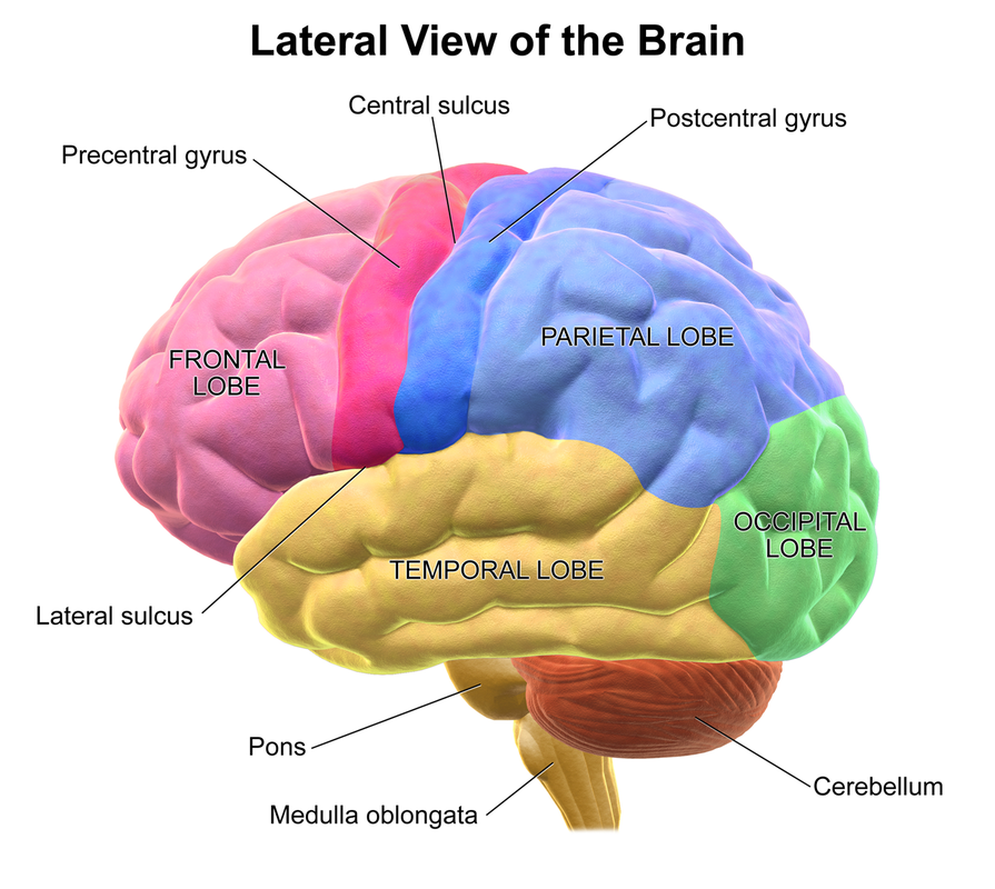
4. Cerebrum: largest portion; envelops the diencephalon; dominant structure; made up of right and left hemispheres connected by the corpus callosum; largest and most complex division of the brain; superior to and surrounding the diencephalon and part of the brain stem; center of higher mental processes and learning, intelligence, communication, memory, reasoning, emotions, interpretation of sensory input, initiation of skeletal muscle contraction; 4 lobes:
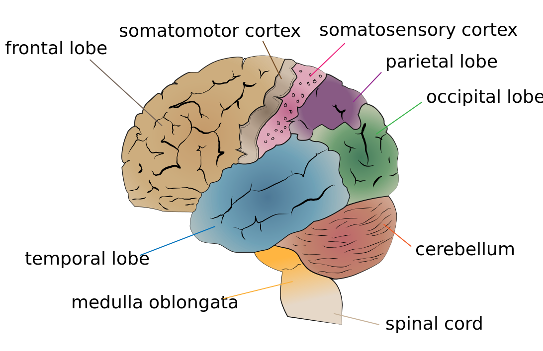
- Frontal lobe
- Parietal lobe
- Temporal lobe
- Occipital lobe
External Anatomical Features:
- Sulci: shallow grooves or furrows between elevations
- Gyri: elevations or folds in the cerebral cortex that increase the surface area for neuron cell bodies
- Central sulcus: shallow goove separating the frontal lobe and the parietal lobe
- Precentral gyrus: elevation located just anterior to the central sulcus
- Postcentral gyrus: elevation located just posterior to the central sulcus
- Longitudinal fissure: deep groove separating the 2 cerebral hemispheres at midline
White Matter: lies deep to the outer cortex and is made mostly of myelinated axons
Corpus Callosum: observable in midsagittal sections of the brain; a band of myelinated axons that connects the 2 cerebral hemispheres
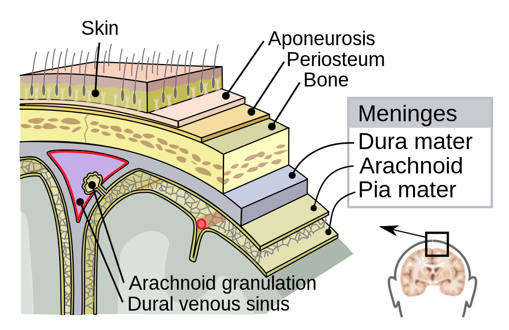
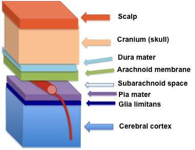
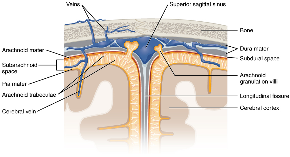
PROTECTION OF THE BRAIN: Cranial Meninges
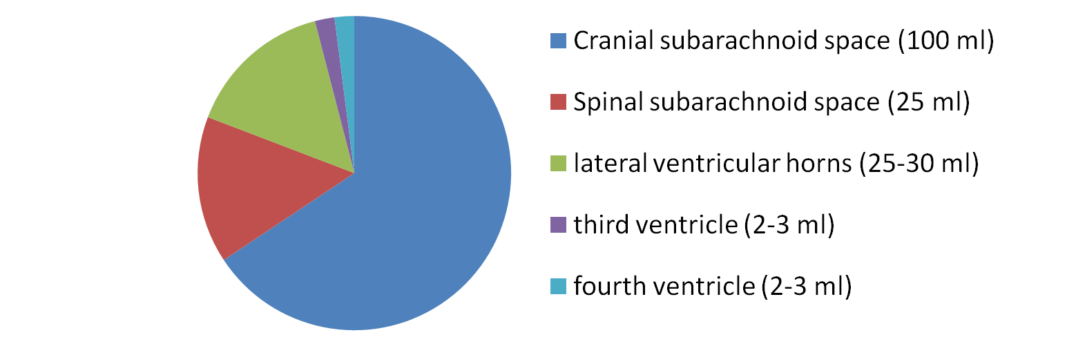
Ventricles: 4 cavities containing choroid plexuses
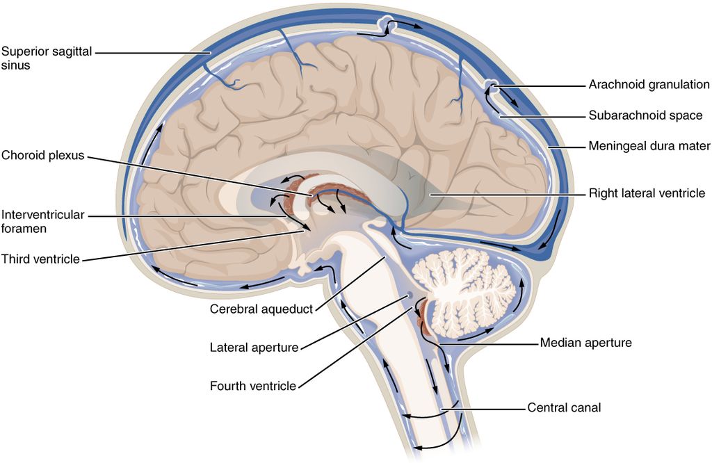
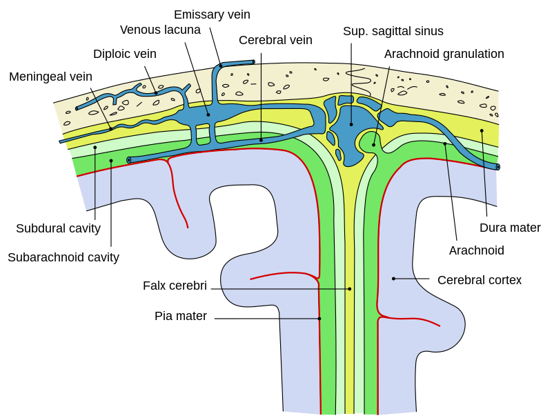
Arachnoid villi: tiny projections of the arachnoid that reabsorb CSF and return it to the blood and into the superior sagittal sinus (a large vein)
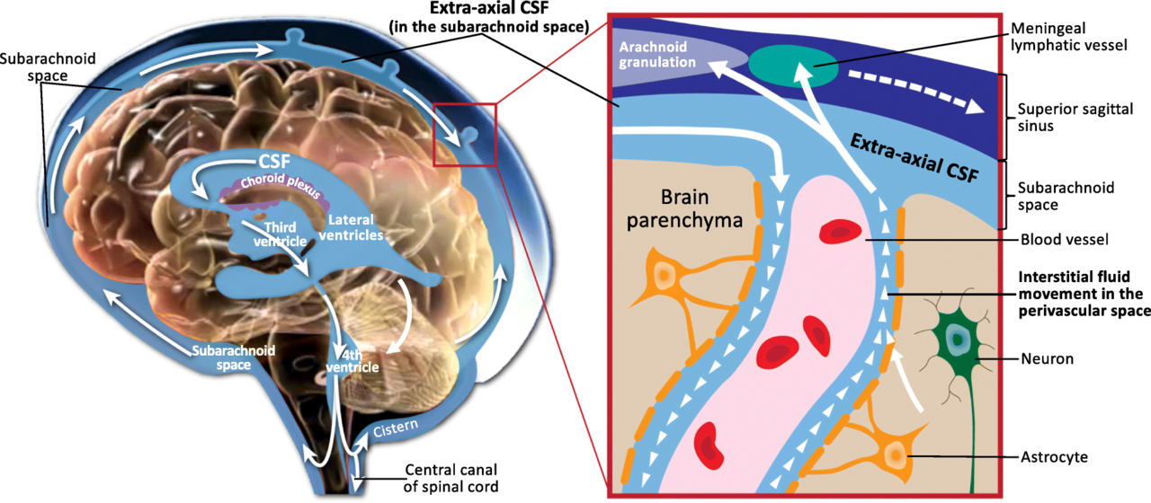
Flow of CSF: 20 mL/hour
- Dura mater (outer)
- Arachnoid mater (middle); spiderweb-like; makes the CSF
- Pia mater (inner)
- Subarachnoid space (between arachnoid and pia mater; contains CSF)
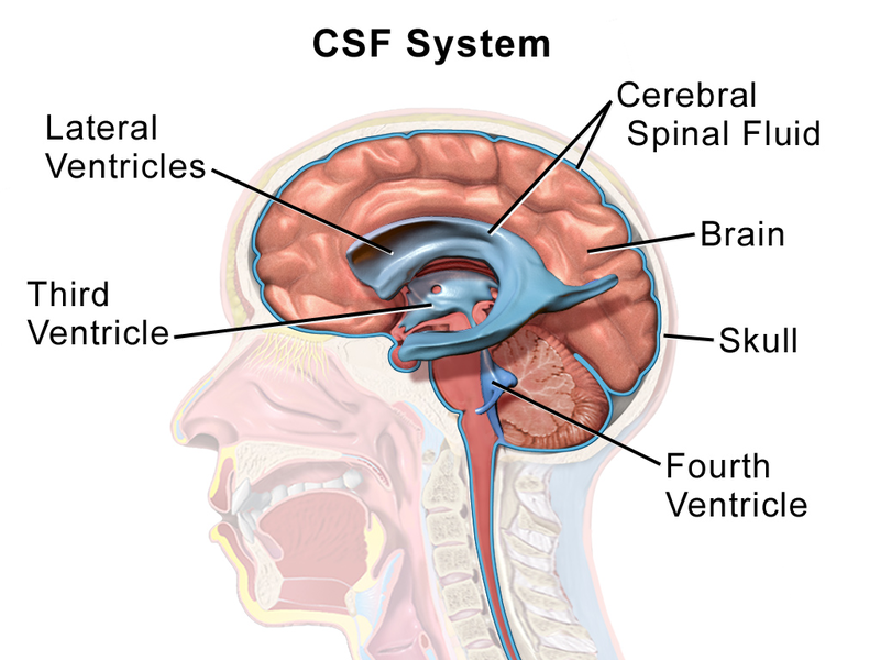
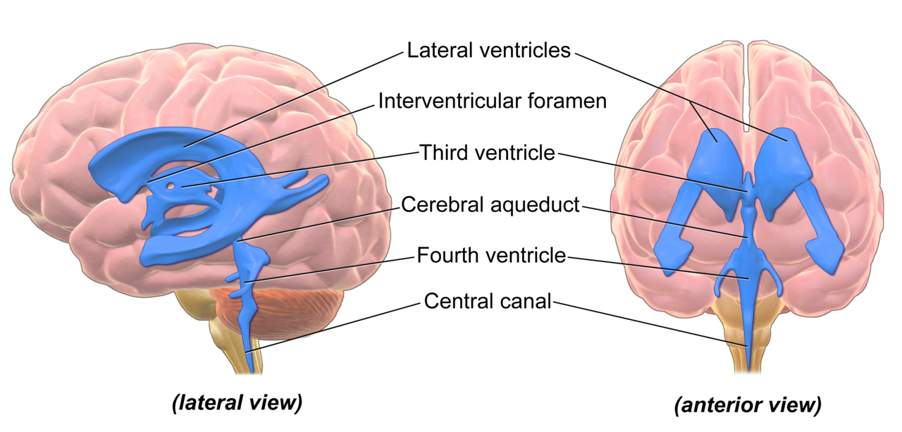
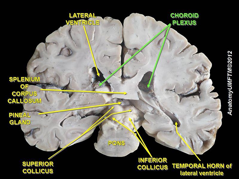
Ventricles: 4 cavities containing choroid plexuses
- Lateral ventricle: located in each cerebral hemisphere; thin membrane separates the 2 ventricles anteriorly; opening connects them to the next ventricle
- Third ventricle: medially located between the paired masses of the thalamus and is narrower and smaller than the other ventricles; connects to the 4th ventricle by a thin tube called the cerebral aqueduct
- Fourth ventricle: last and final ventricle located between the pons and the cerebellum; openings allow CSF to flow into the subarachnoid space surrounding the brain and spinal cord
Arachnoid villi: tiny projections of the arachnoid that reabsorb CSF and return it to the blood and into the superior sagittal sinus (a large vein)
Flow of CSF: 20 mL/hour
- Choroid Plexuses
- Lateral ventricles
- Third ventricle
- Fourth ventricle
- Cerebral aqueduct (canal)
- Subarachnoid space
- Central canal
- Epidural space
- Arachnoid villi (reabsorption)
- Superior sagittal sinus (vein)
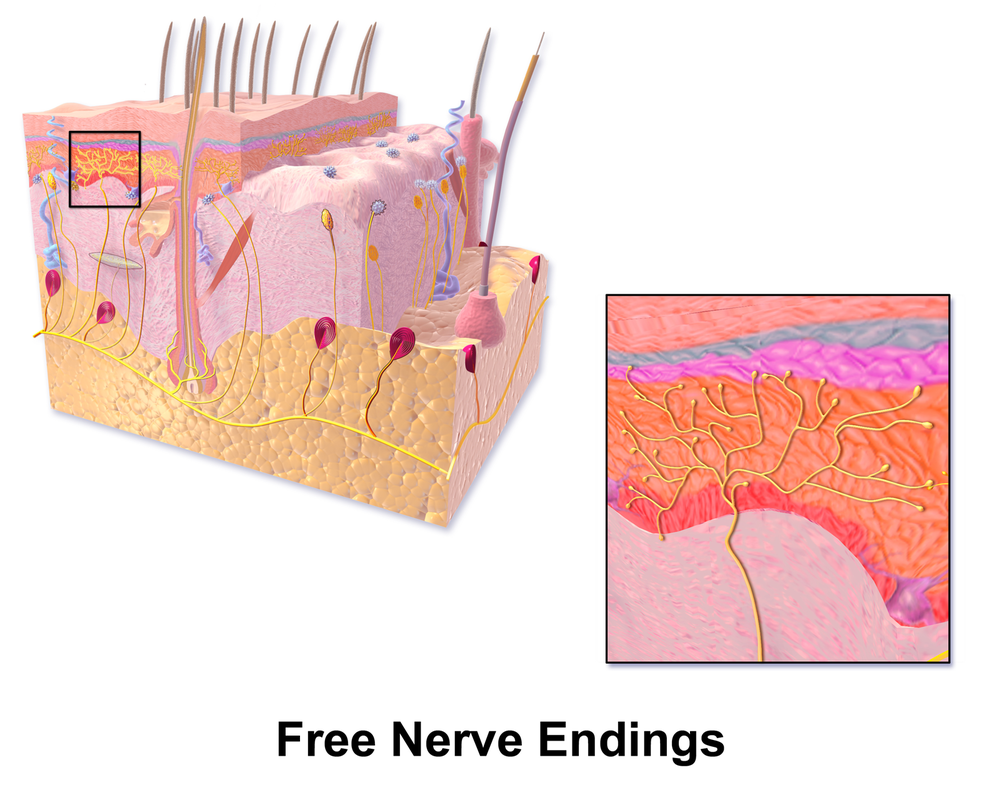
Free Nerve Endings:
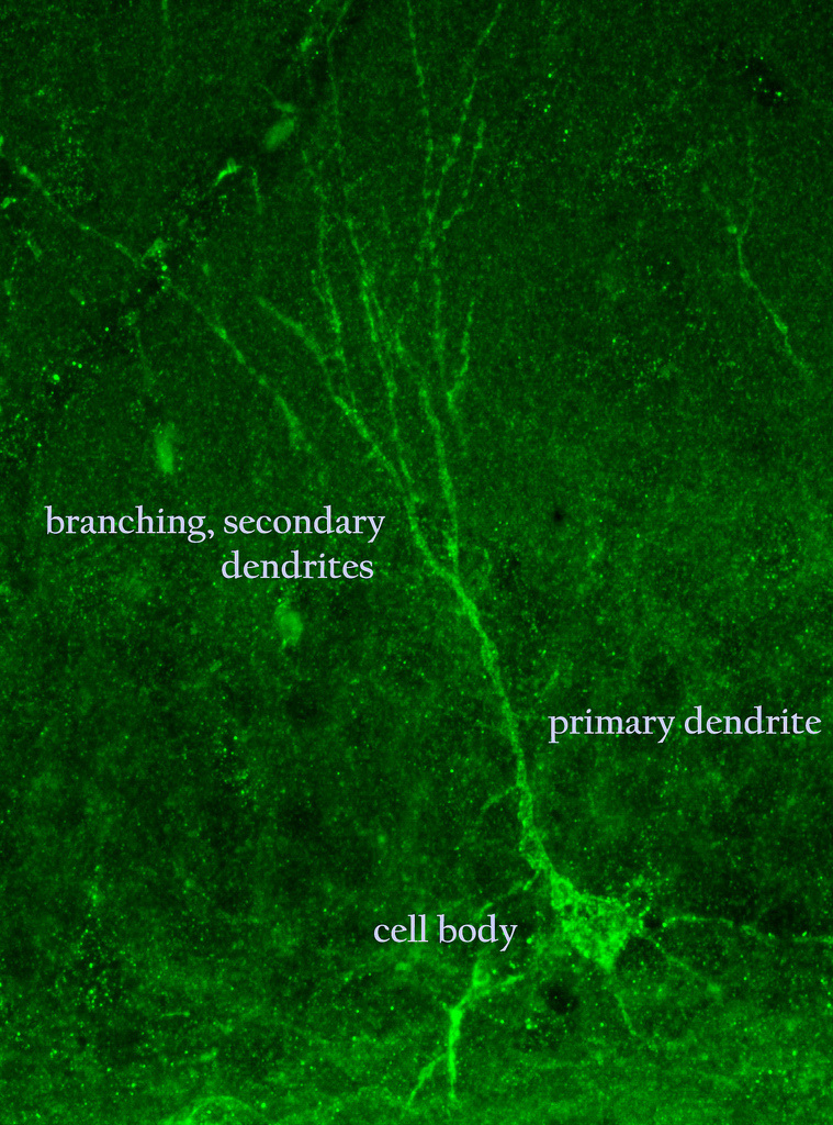
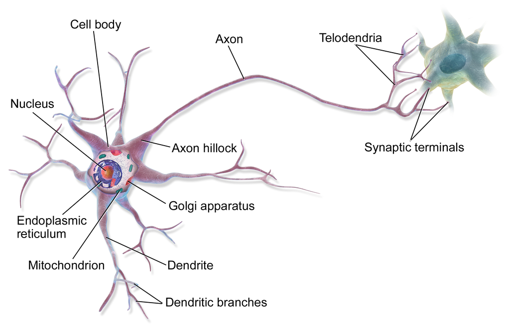
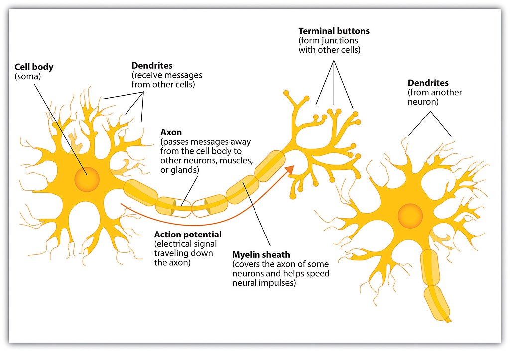
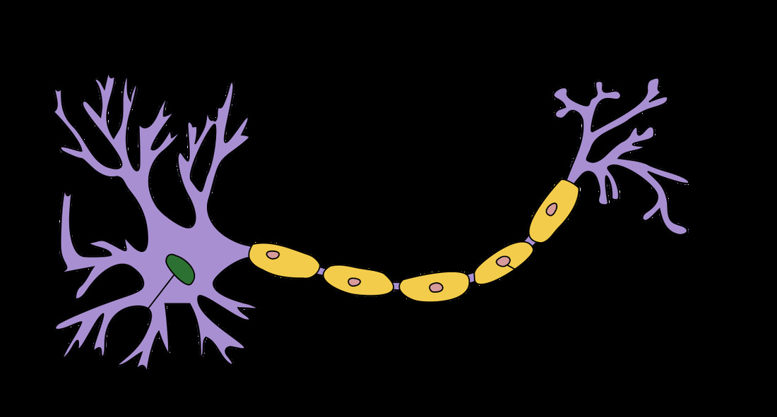
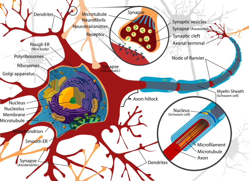
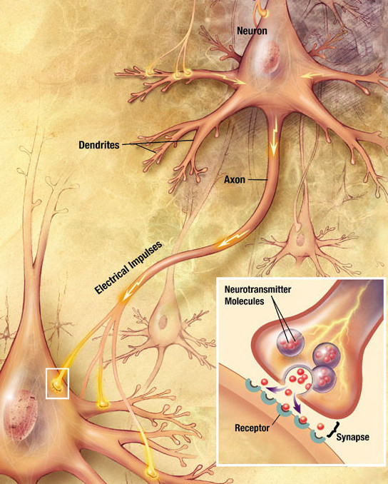
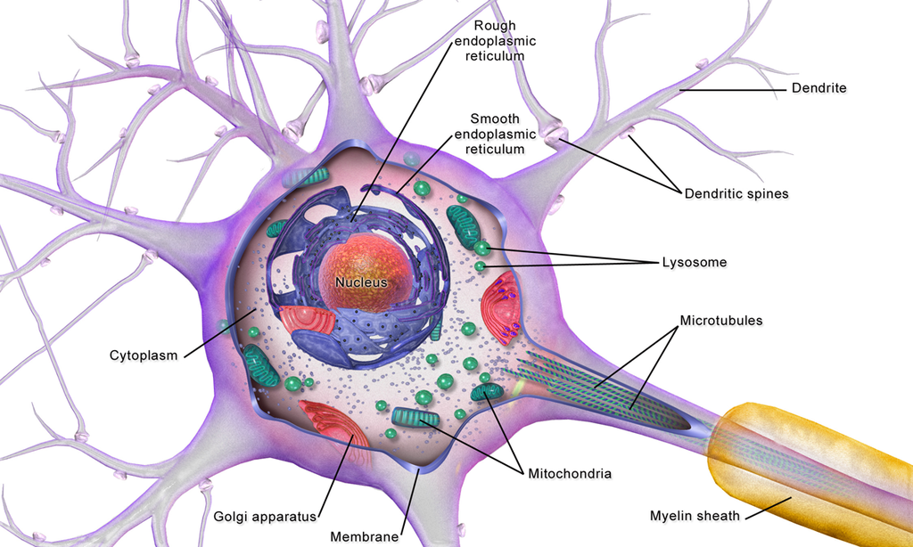
The Structure of a Nerve Cell:
By BruceBlaus - Own work, CC BY 3.0, https://commons.wikimedia.org/w/index.php?curid=28761830
By Jennifer Walinga - https://opentextbc.ca/introductiontopsychology/chapter/3-1-the-neuron-is-the-building-block-of-the-nervous-system/, CC BY-SA 4.0, https://commons.wikimedia.org/w/index.php?curid=97847412
By BruceBlaus - Own work, CC BY-SA 4.0, https://commons.wikimedia.org/w/index.php?curid=46621398
By https://www.scientificanimations.com/ - http://www.scientificanimations.com/wiki-images/, CC BY-SA 4.0, https://commons.wikimedia.org/w/index.php?curid=72340357
By Doctor Jana - https://docjana.com/neuro-muscular-junction/, CC BY 4.0, https://commons.wikimedia.org/w/index.php?curid=46835961
Thomas Splettstoesser (www.scistyle.com), CC BY-SA 4.0 , via Wikimedia Commons
By Isaac Oster - Own work, CC BY-SA 4.0, https://commons.wikimedia.org/w/index.php?curid=69565964
By Doctor Jana - http://docjana.com/#/gbs; http://www.patreon.com/posts/guillain-barre-4374004, CC BY 4.0, https://commons.wikimedia.org/w/index.php?curid=46847816
Nerves Throughout the Body:
By Neuron_with_oligodendrocyte_and_myelin_sheath.svg: *Complete_neuron_cell_diagram_en.svg: LadyofHatsderivative work: Andrew c (talk) - Neuron_with_oligodendrocyte_and_myelin_sheath.svg, Public Domain, https://commons.wikimedia.org/w/index.php?curid=10888009
|
OpenStax, CC BY 4.0
|
By Dchordpdx - Own work, CC BY 4.0, https://commons.wikimedia.org/w/index.php?curid=64427063
By Archontia Kaminari - Own work, CC BY-SA 4.0, https://commons.wikimedia.org/w/index.php?curid=45342606
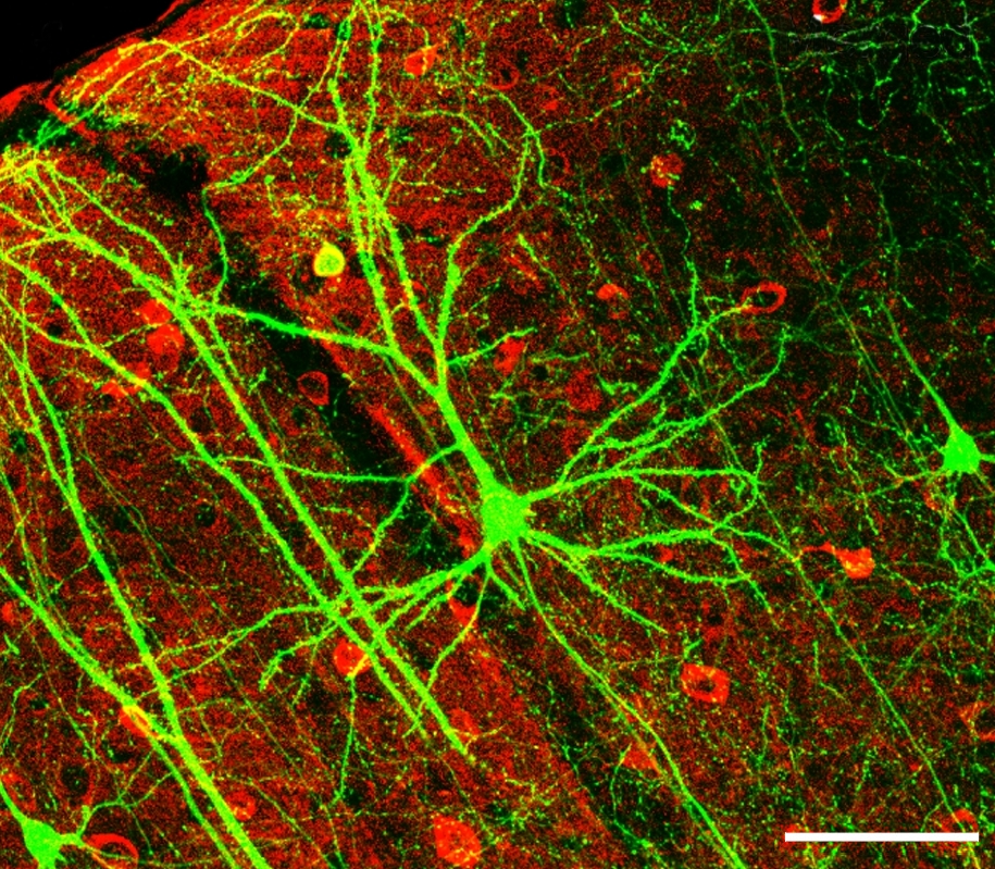
https://upload.wikimedia.org/wikipedia/commons/0/0b/Microglia_and_neurons.jpg
GerryShaw, CC BY-SA 3.0 , via Wikimedia Commons
https://upload.wikimedia.org/wikipedia/commons/2/2e/Microglial_cells_%28red%29_in_rat_cerebellar_molecular_layer.jpg
GerryShaw, CC BY-SA 4.0 , via Wikimedia Commons
By J. Wegiel - Research, CC BY 4.0, https://commons.wikimedia.org/w/index.php?curid=83491274
By Artwork by Holly Fischer - http://open.umich.edu/education/med/resources/second-look-series/materials - CNS Slide 4, CC BY 3.0, https://commons.wikimedia.org/w/index.php?curid=24367125
|
By ArizonaLifeScience - Own work Orchinik Lab, Arizona State University, Public Domain, https://commons.wikimedia.org/w/index.php?curid=10018500
|
By BruceBlaus. When using this image in external sources it can be cited as:Blausen.com staff (2014). "Medical gallery of Blausen Medical 2014". WikiJournal of Medicine 1 (2). DOI:10.15347/wjm/2014.010. ISSN 2002-4436. - Own work, CC BY 3.0, https://commons.wikimedia.org/w/index.php?curid=28761843
By Nephron - Own work, CC BY-SA 3.0, https://commons.wikimedia.org/w/index.php?curid=12108539
Autonomic Nervous System:
By Geo-Science-International - Own work, CC0, https://commons.wikimedia.org/w/index.php?curid=47377075
By OpenStax College - Anatomy & Physiology, Connexions Web site. http://cnx.org/content/col11496/1.6/, Jun 19, 2013., CC BY 3.0, https://commons.wikimedia.org/w/index.php?curid=30148020
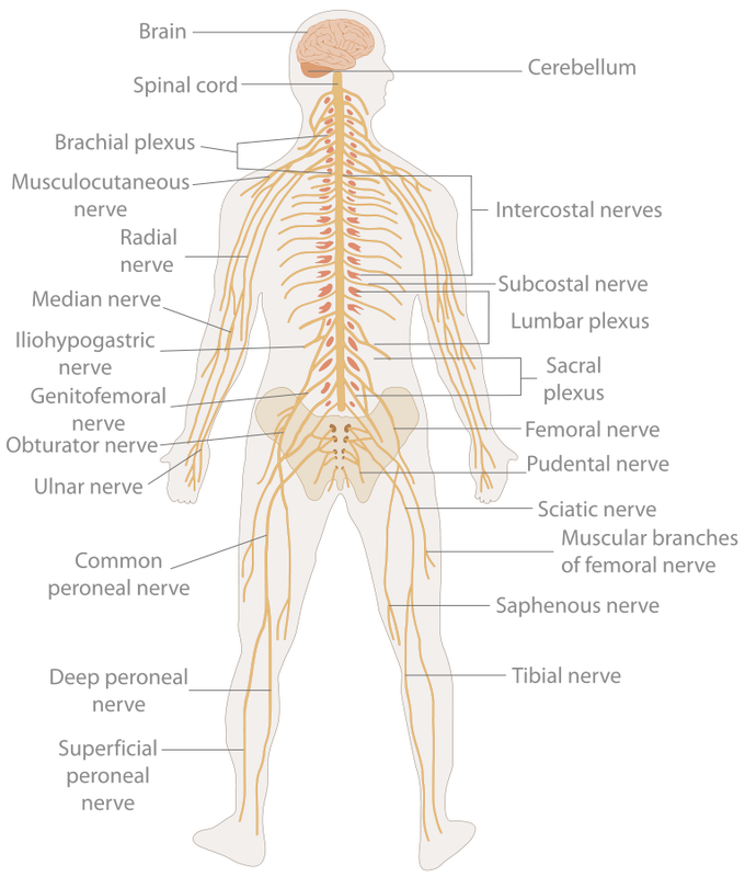
Peripheral Nervous System:
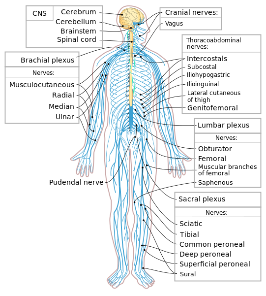
By This SVG image was created by Medium69.Cette image SVG a été créée par Medium69.Please credit this : William Crochot - File:Nervous system diagram.png, CC BY-SA 4.0, https://commons.wikimedia.org/w/index.php?curid=36395693
By Cancer Research UK - Original email from CRUK, CC BY-SA 4.0, https://commons.wikimedia.org/w/index.php?curid=34332941
|
By Henry Vandyke Carter - Vertebral column image.- From: Henry Gray (1918) Anatomy of the Human Body (See "Book" section below)- Altered by User:Uwe Gille, Public Domain, https://commons.wikimedia.org/w/index.php?curid=1282158
|
By Tomwsulcer - Own work, CC0, https://commons.wikimedia.org/w/index.php?curid=15006027
|
By Polarlys and Mikael Häggström - File:Medulla spinalis - tracts - English.svg by Polarlys (translation by Selket)., CC BY-SA 3.0, https://commons.wikimedia.org/w/index.php?curid=10909281
|
By BruceBlaus - Own work, CC BY-SA 4.0, https://commons.wikimedia.org/w/index.php?curid=46621399
By user:debivort - Own work, CC BY-SA 3.0, https://commons.wikimedia.org/w/index.php?curid=1675049
By Dr. Johannes Sobotta - Sobotta's Textbook and Atlas of Human Anatomy 1908, Public Domain, https://commons.wikimedia.org/w/index.php?curid=29190153
By Dr. Johannes Sobotta - Sobotta's Textbook and Atlas of Human Anatomy 1908, Public Domain, https://commons.wikimedia.org/w/index.php?curid=29190149
By Dr. Johannes Sobotta - Sobotta's Textbook and Atlas of Human Anatomy 1908, Public Domain, https://commons.wikimedia.org/w/index.php?curid=29190151
By Zuzanna K. Filutowska - Own work, CC BY-SA 3.0, https://commons.wikimedia.org/w/index.php?curid=27591940
By Z22 - Own work, CC BY-SA 4.0, https://commons.wikimedia.org/w/index.php?curid=38767462
By Anatomist90 - Own work, CC BY-SA 3.0, https://commons.wikimedia.org/w/index.php?curid=26137757
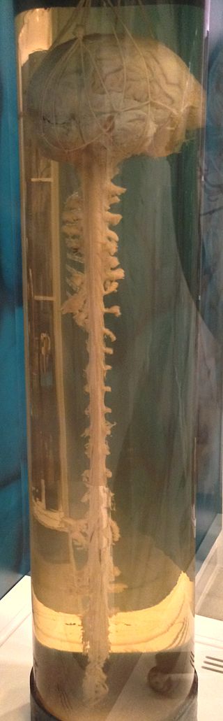
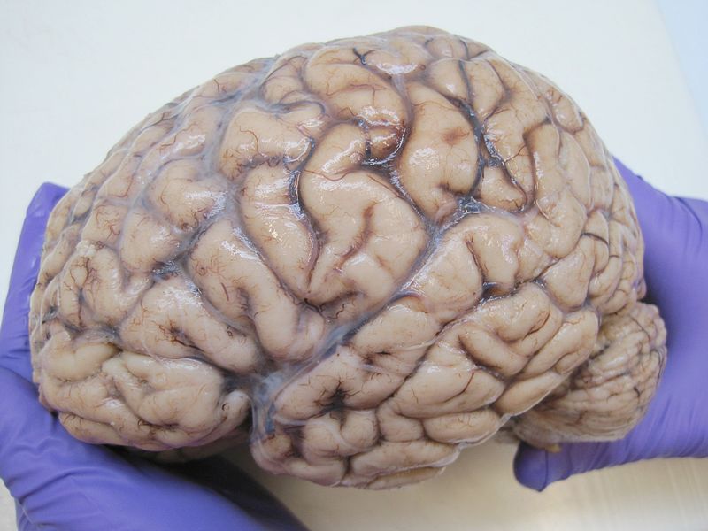
The Brain:
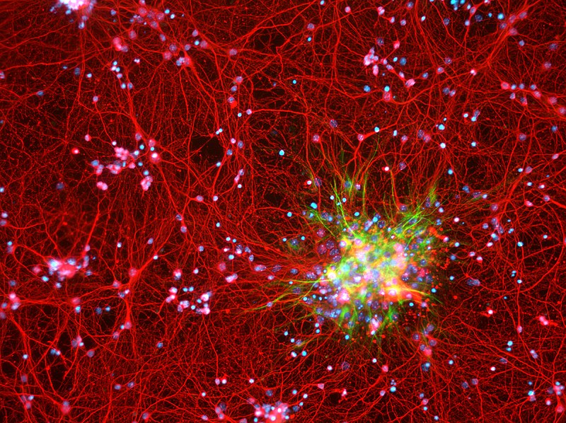
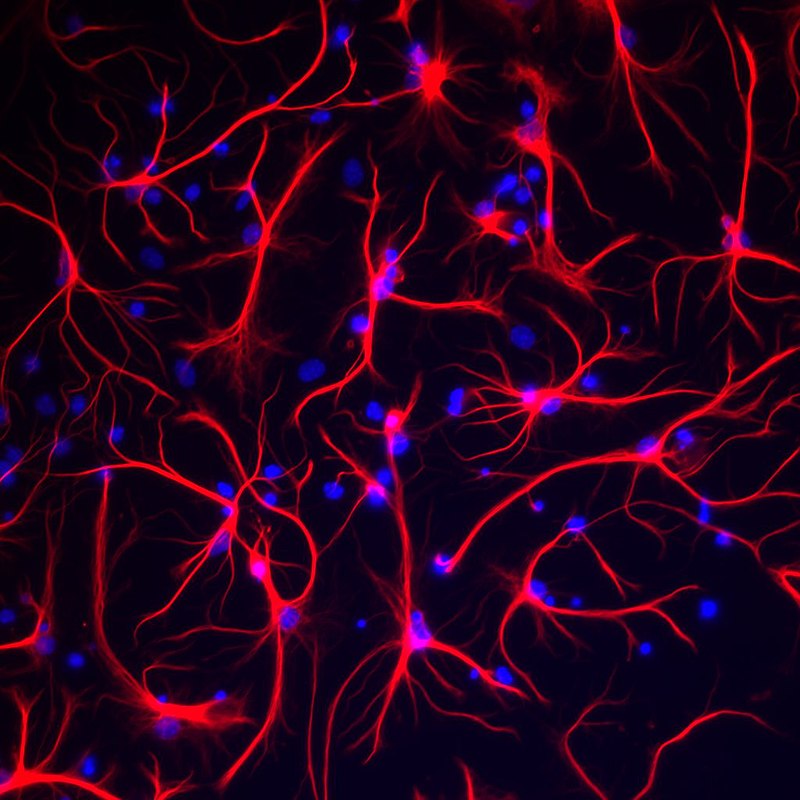
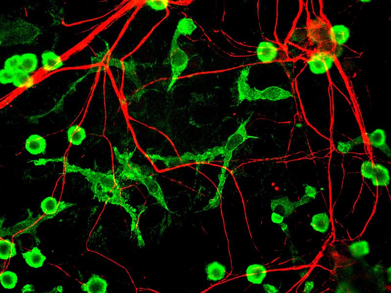
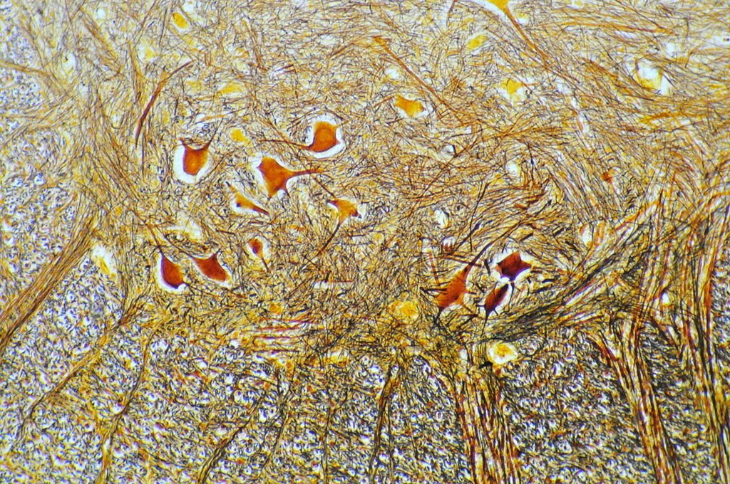
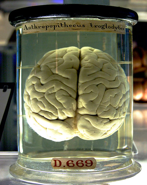
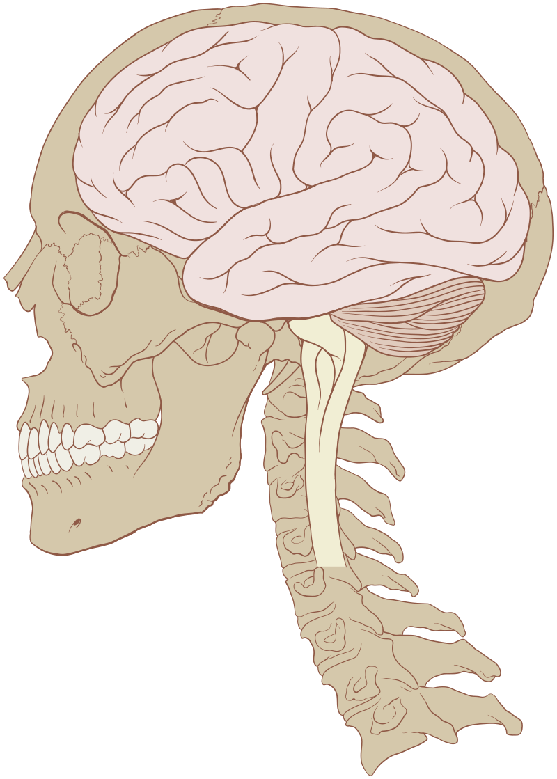
The brain is the organ that is the control center of the central nervous system. It is the most complex organ and it is protected by the skull bones. The outer portion of the brain is called the cerebrum, or the cerebral cortex, and it contains anywhere from 15 billion to 33 billion neurons of various types as well as glial (support) cells. See the neurons above for more information.
The brain controls the other organs of the body. It sends signals called neurotransmitters and hormones that tell the body what to do and aids in the coordination of responses to stimuli. Inside, the brain consists of darker colored grey matter, unmyelinated cells, and lighter colored white matter, myelinated cells that help send signals along faster from nerve cell to nerve cell.
The brain controls the other organs of the body. It sends signals called neurotransmitters and hormones that tell the body what to do and aids in the coordination of responses to stimuli. Inside, the brain consists of darker colored grey matter, unmyelinated cells, and lighter colored white matter, myelinated cells that help send signals along faster from nerve cell to nerve cell.
By Jensflorian - Own work, CC BY-SA 4.0, https://commons.wikimedia.org/w/index.php?curid=39237155
By Anatomist90 - Own work, CC BY-SA 3.0, https://commons.wikimedia.org/w/index.php?curid=19132692
By Anatomist90 - Own work, CC BY-SA 3.0, https://commons.wikimedia.org/w/index.php?curid=29071566
By Gaetan Lee . Tilt corrected by Kaldari. - originally posted to Flickr as Chimp Brain in a jar, CC BY 2.0, https://commons.wikimedia.org/w/index.php?curid=28819747
By Patrick J. Lynch, medical illustrator - Patrick J. Lynch, medical illustrator, CC BY 2.5, https://commons.wikimedia.org/w/index.php?curid=1496706
|
By Dr. Johannes Sobotta - Atlas and Text-book of Human Anatomy Volume III Vascular System, Lymphatic system, Nervous system and Sense Organs, Public Domain, https://commons.wikimedia.org/w/index.php?curid=29135452
|
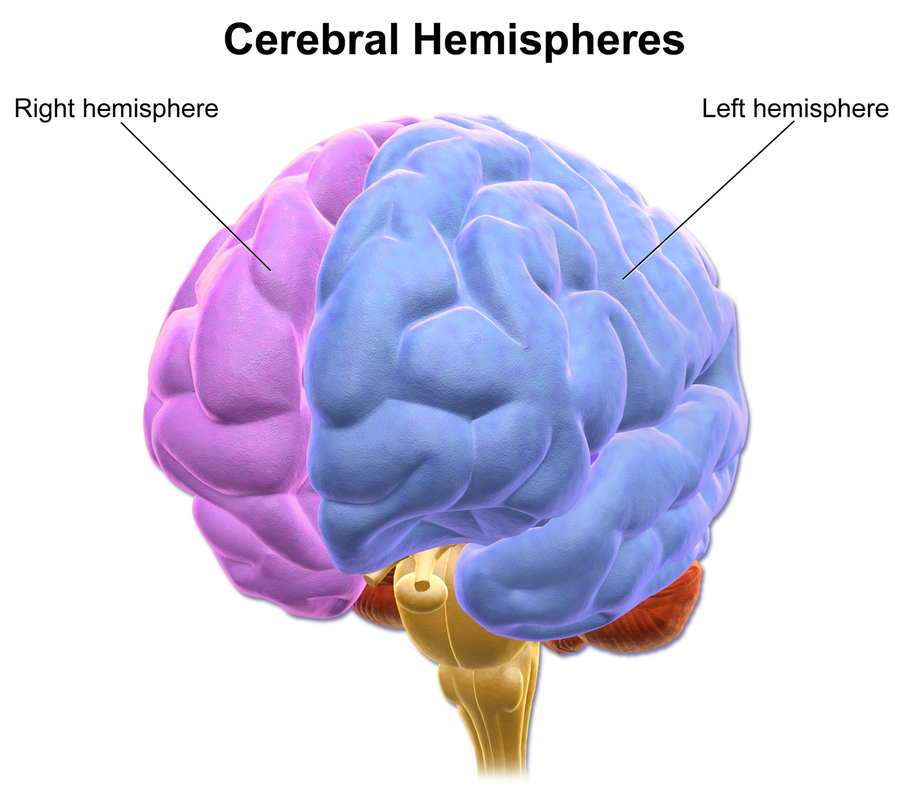
The brain is divided into 2 cerebral hemispheres: the right and left hemispheres, separated by a long groove called the longitudinal fissure. The 2 hemispheres are linked inside the brain by a structure known as the corpus callosum, which consists of a huge bundle of nerve fibers for communication between the 2 hemispheres.
- Near mirror images of each other
- Right hemisphere has a slight warping known as the Yakovlevian torque
- Receives information from opposite sides of the body, respectively
By BruceBlaus. When using this image in external sources it can be cited as:Blausen.com staff (2014). "Medical gallery of Blausen Medical 2014". WikiJournal of Medicine 1 (2). DOI:10.15347/wjm/2014.010. ISSN 2002-4436. - Own work, CC BY 3.0, https://commons.wikimedia.org/w/index.php?curid=31118596
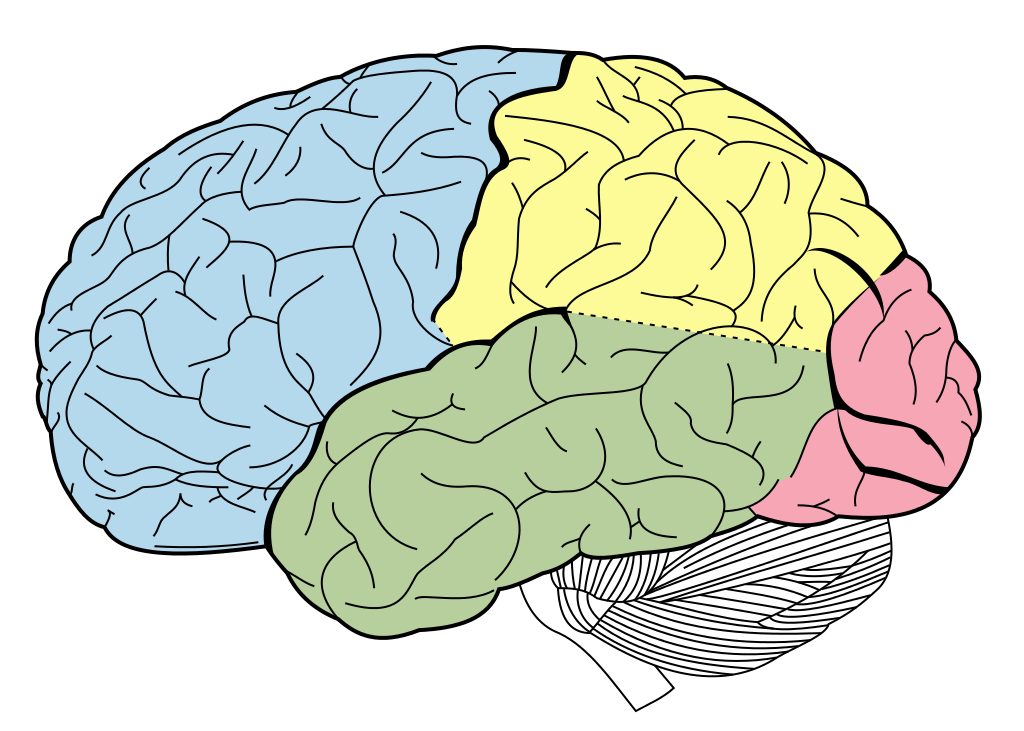
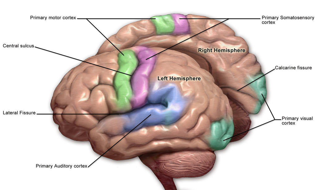
The cerebral cortex also consists of specific lobes:
- Frontal Lobe
- Central Sulcus-prominant fissure/groove that separates the frontal and parietal lobes; separates the primary motor cortex from the primary somatosensory cortex
- Precentral Gyrus
- Central Sulcus-prominant fissure/groove that separates the frontal and parietal lobes; separates the primary motor cortex from the primary somatosensory cortex
- Parietal Lobe
- Postcentral Gyrus
- Postcentral Gyrus
- Occipital Lobe
- Temporal Lobe
By BruceBlaus - Own work, CC BY 3.0, https://commons.wikimedia.org/w/index.php?curid=31118589
The 3 major parts of the outer brain include:
a) The cerebrum or cerebral cortex (outer)
b) The cerebellum
c) The brainstem
a) The cerebrum or cerebral cortex (outer)
b) The cerebellum
c) The brainstem
The Cerebrum/Cerebral Cortex:
- The outer brain layer consisting of grey matter, which is 2-4 mm thick
- Mostly cell bodies
- Mostly astrocytes
- Capillaries
- Mostly cell bodies
- Consists of the 2 hemispheres
- Consists of the 4 lobes
- Consists of fissures/grooves and convolutions and folds
- Gyrus/gyri: folds/ridges
- Sulcus/sulci: fissures/grooves
- Gyrus/gyri: folds/ridges
- Interior consists of inner core of white matter (centrum semiovale)
- Lateral ventricles
- Basal nuclei
- White matter
- Myelinated sheaths of neuronal axons
- Lateral ventricles
- Neocortex is made up of 6 layers (51 different areas) and each region is known as Brodmann areas, which play a role in sensation, cognition and behavior
- Plays a key role in the following:
- Memory
- Attention
- Perception
- Awareness
- Thought
- Language (speech, processing, understanding)
- Consciousness
- Memory
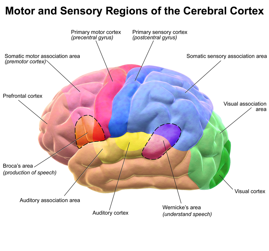
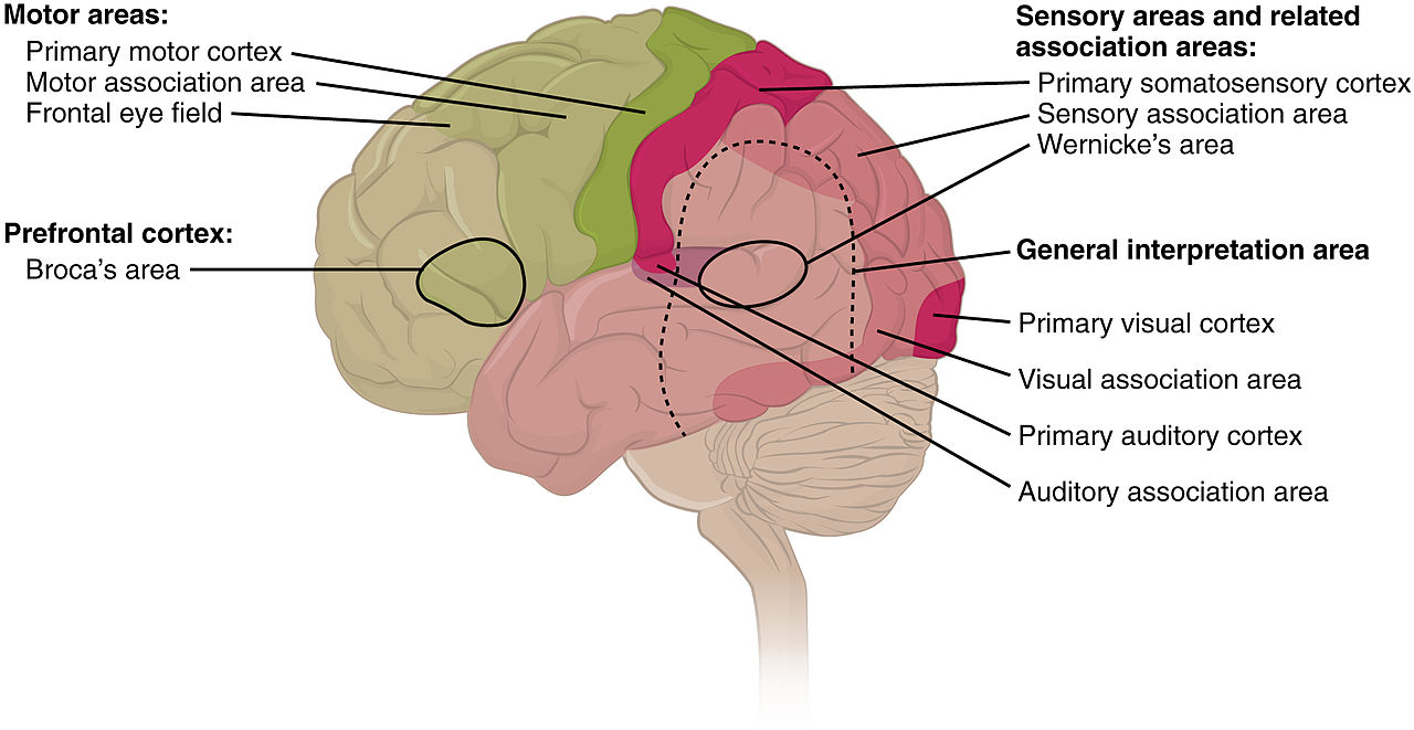
Cerebral Cortex Areas and Functions:
I. Sensory Areas:
- Primary Sensory Areas: receive and process information from the senses by receipt of sensory input from the thalamus
- Primary Visual Cortex: senses of vision
- Primary Auditory Cortex: senses of hearing
- Primary Somatosensory Cortex: senses of touch
- Primary Visual Cortex: senses of vision
- Primary Motor Areas: control voluntary movement
- Secondary Motor Areas and Premotor Cortex: select voluntary movement
- Posterior Parietal Cortex: voluntary movements in space
- Dorsolateral Premotor Cortex: decides which movements to take to higher order instructions, more precision
By BruceBlaus. When using this image in external sources it can be cited as:Blausen.com staff (2014). "Medical gallery of Blausen Medical 2014". WikiJournal of Medicine 1 (2). DOI:10.15347/wjm/2014.010. ISSN 2002-4436. - Own work, CC BY 3.0, https://commons.wikimedia.org/w/index.php?curid=31574257
By OpenStax College - Anatomy & Physiology, Connexions Web site. http://cnx.org/content/col11496/1.6/, Jun 19, 2013., CC BY 3.0, https://commons.wikimedia.org/w/index.php?curid=30148119
Basal Ganglia:
- Lie just underneath the cerebral cortex
- Masses of grey matter
- Receive sensory input from the midbrain and from certain areas of the motor cortex of the cerebrum
The Cerebellum:
- The bottom portion of the brain
- Controls balance
- Controls other brain systems and makes them precise
- Without it, we would be hesitant and clumsy
- Controls muscle coordination and is responsible for the precise movements and timing of movements of all voluntary muscles of the body
- Half of all the brain's neurons are located there
- Participates in motor learning of adjustments and parameters of movement
The Brainstem:
Consists of 3 parts:
a) Midbrain
a) Midbrain
- There are motor neurons here called oculomotor nuclei that directly stimulate the eye muscles
- Coordination of movements of arms and legs
- Involved in sleep-wake cycle and alertness
- Contains nuclei
- Involved in many voluntary functions
- Controls things like sleep, respiration, swallowing, bladder function, equilibrium, eye movement, facial expressions, posture, walking, breathing
- Involved in sleep-wake cycle and alertness
- Contains many small nuclei
- Involved in many sensory and involuntary motor functions, including vomiting, digestive processes, and heart rate
|
|
By Dr. Johannes Sobotta - Atlas and Text-book of Human Anatomy Volume III Vascular System, Lymphatic system, Nervous system and Sense Organs, Public Domain, https://commons.wikimedia.org/w/index.php?curid=29135453
By BruceBlaus. When using this image in external sources it can be cited as:Blausen.com staff (2014). "Medical gallery of Blausen Medical 2014". WikiJournal of Medicine 1 (2). DOI:10.15347/wjm/2014.010. ISSN 2002-4436. - Own work, CC BY 3.0, https://commons.wikimedia.org/w/index.php?curid=31118595
By OpenStax - https://cnx.org/contents/[email protected]:fEI3C8Ot@10/Preface, CC BY 4.0, https://commons.wikimedia.org/w/index.php?curid=30147960
By BruceBlaus - Own work, CC BY 3.0, https://commons.wikimedia.org/w/index.php?curid=28761845
By Mark D. Shen - Shen MD. Cerebrospinal fluid and the early brain development of autism. J Neurodev Disord. 2018;10(1):39. Published 2018 Dec 13. https://dx.doi.org/10.1186%2Fs11689-018-9256-7, CC BY 4.0, https://commons.wikimedia.org/w/index.php?curid=79736902
By Mysid - Made by Mysid Inkscape, based on plate 769 from Gray's Anatomy (1918, public domain)., Public Domain, https://commons.wikimedia.org/w/index.php?curid=10493338
By Anatomist90 - Own work, CC BY-SA 3.0, https://commons.wikimedia.org/w/index.php?curid=19132819
By User:TarsaucerUploaded by Tarsaucer at en.wikipedia - Author, CC0, https://commons.wikimedia.org/w/index.php?curid=16844162
By BruceBlaus. When using this image in external sources it can be cited as:Blausen.com staff (2014). "Medical gallery of Blausen Medical 2014". WikiJournal of Medicine 1 (2). DOI:10.15347/wjm/2014.010. ISSN 2002-4436. - Own work, CC BY 3.0, https://commons.wikimedia.org/w/index.php?curid=29987039
By BruceBlaus. When using this image in external sources it can be cited as:Blausen.com staff (2014). "Medical gallery of Blausen Medical 2014". WikiJournal of Medicine 1 (2). DOI:10.15347/wjm/2014.010. ISSN 2002-4436. - Own work, CC BY 3.0, https://commons.wikimedia.org/w/index.php?curid=27796935
By James Heilman, MD - Own work, CC BY 3.0, https://commons.wikimedia.org/w/index.php?curid=6992139
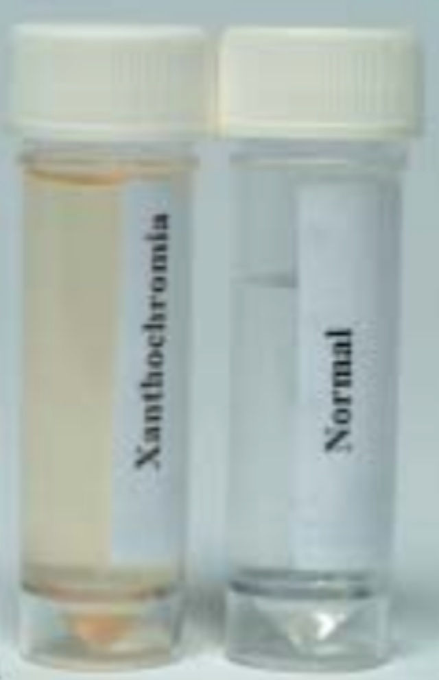
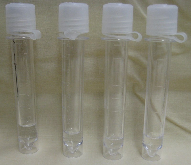
Xanthochromic CSF is "yellowish" in color rather than clear and colorless.
https://www.scientificanimations.com, CC BY-SA 4.0 , via Wikimedia Commons
By SVG by Mysid, original by SEER Development Team [1], Jmarchn - Vectorized in Inkscape by Mysid, based on work by SEER Development Team, CC BY-SA 3.0, https://commons.wikimedia.org/w/index.php?curid=10485059
By Katie Ahlers - I created this through powerpoint., CC BY-SA 4.0, https://commons.wikimedia.org/w/index.php?curid=14693493
By OpenStax - https://cnx.org/contents/[email protected]:fEI3C8Ot@10/Preface, CC BY 4.0, https://commons.wikimedia.org/w/index.php?curid=30147959
By Mysid - Made by Mysid Inkscape, based on plate 770 from Gray's Anatomy (1918, public domain)., Public Domain, https://commons.wikimedia.org/w/index.php?curid=10496507
By Brain_human_normal_inferior_view_with_labels_en.svg: *Brain_human_normal_inferior_view.svg: Patrick J. Lynch, medical illustratorderivative work: Beaoderivative work: Dwstultz (talk) - Brain_human_normal_inferior_view_with_labels_en.svg, CC BY 2.5, https://commons.wikimedia.org/w/index.php?curid=15108118
Dental: anesthesia, numbing the nerves to decrease pain for dental procedures in the mouth
Radiography: X-rays/scans of the brain, bones, other structures will help you see brain tumors, bone breaks where nerve damage has occurred, will help the anesthesiologist know where to numb certain nerves, see signs of stroke, dementia, Parkinson's disease, Alzheimer's disease, blockages, traumatic injuries, microinjuries, TIA's, hematomas, and other potential things that could save someone's life
Ultrasound: this helps check for nerve function, helps physicians with guided needle biopsies so that they miss hitting or damaging nerves in the process, view blood clots that may be putting pressure on or causing damage to nerves nearby
Massage Therapy: people come in pain, and your role is to help reduce pain levels and increase range of motion in conjunction with some other providers, so you are helping to release neurotransmitters called endorphins and enkaphalins to aid in relaxation of muscles and nerves
Surgical Tech: you are assisting with surgeries and may even be assisting with the administration of neurotransmitters before surgery, which cause numbness and put the nerves "to sleep" so repair can take place (surgical)
MA/PA/Nurses: You are looking for causes of and diagnosis of causes of pain so you can administer treatment either under the guidance of a doctor, or you will be the leader of this if you are a doctor yourself or a PA or NP. You may be administering medications that act on nerves: norepinephrine/epinephrine in EPI-pens to stop an anaphylactic allergic reaction, medications containing antihistamine to block release of allergy-causing histamine, prostaglandin as a hormone treatment, anesthesia for surgery or for numbing (think about even Lidocaine...), melatonin for kids having difficulty sleeping, dopamine for those with Parkinson's disease, acetylcholine to treat M.S. or Myasthenia gravis
H.I.T.: Terminology! You will use all of this key terminology every day in your role of entering and extracting information, keeping track of patient records, analyzing them for accuracy, etc...
