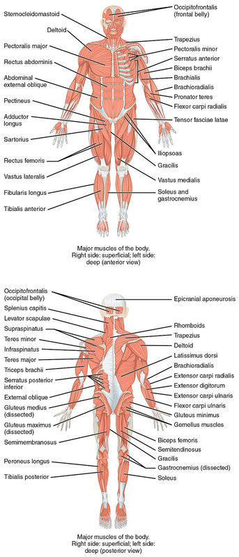
MAJOR MUSCLES OF THE BODY:
Lecture Objectives: Muscular System
Upon completion of this chapter and lectures, you should be able to:
Lab Objectives: Exercises
Upon completion of these exercises, you should be able to:
Upon completion of this chapter and lectures, you should be able to:
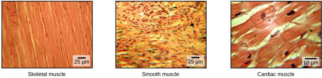
- List, locate in the body, and compare the structure and function of the three major types of muscle tissue.
- Discuss the microscopic structure and function of a skeletal muscle, including the sarcomere and motor unit.
- Discuss how a muscle is stimulated and compare the major types of skeletal muscle contractions.
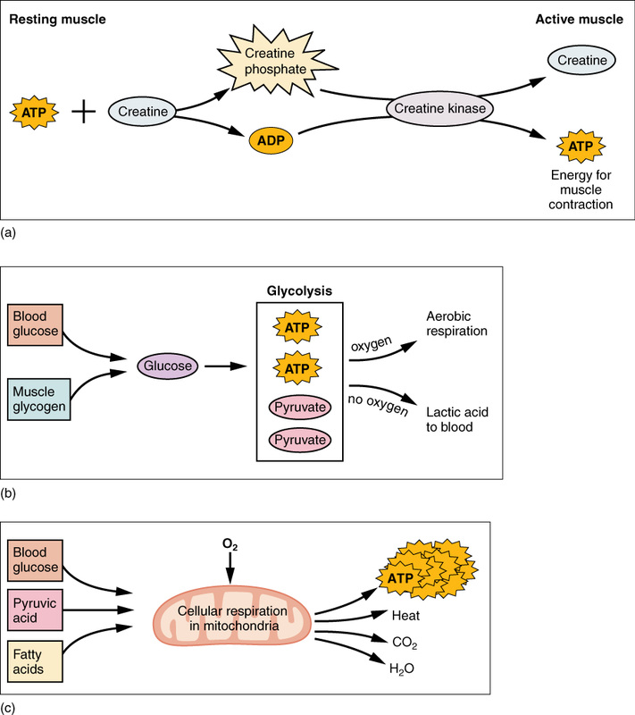
- List and explain the primary effects of exercise on the structure and function of skeletal muscles.
- List and explain the most common types of movement produced by skeletal muscles.
- Name, identify on a model or diagram, and give the function of the major muscles of the body discussed in this chapter.
Lab Objectives: Exercises
Upon completion of these exercises, you should be able to:
- Describe the function of the connective tissue coverings of skeletal muscles.
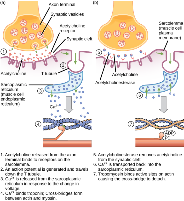
- Describe the structure of the neuromuscular junction.
- Describe how skeletal muscles achieve a smooth, sustained contraction.
- Define threshold of contraction, maximal stimulus, recruitment, and fatigue, and explain how to observe them.
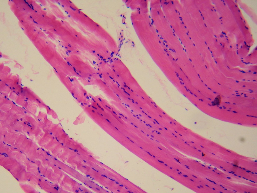
- Identify longitudinal and cross-sections of muscle tissues.
- Describe how skeletal muscles are named.
- Identify major skeletal muscles on models or illustrations.
- Describe the actions of the major skeletal muscles.
https://knowledge.carolina.com/discipline/life-science/biology/chicken-wing-musculature/
THE MUSCULOSKELETAL SYSTEM AND MUSCLES:
SKELETAL MUSCLE CELLS AND FIBERS:
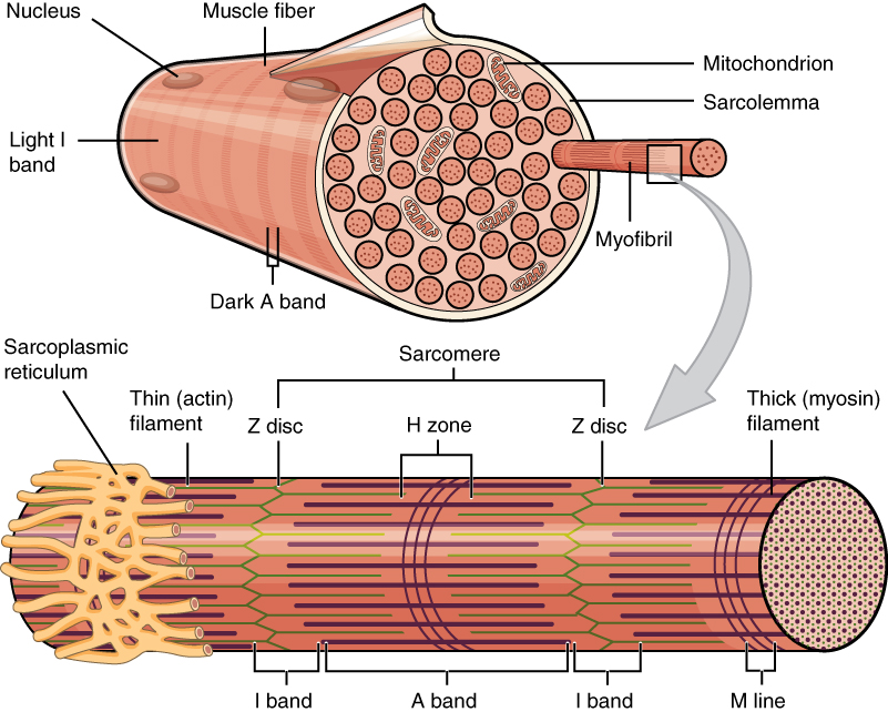
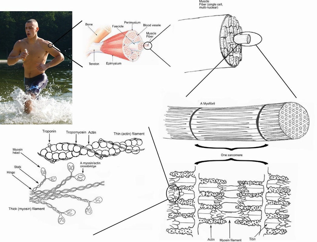
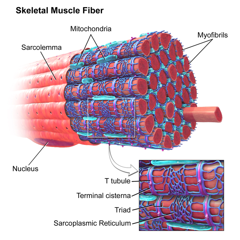
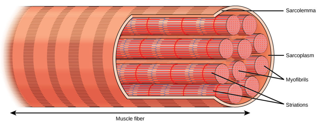
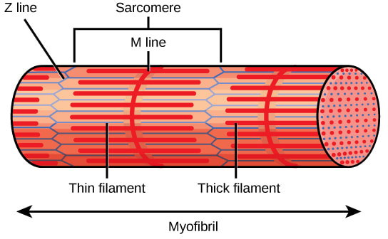
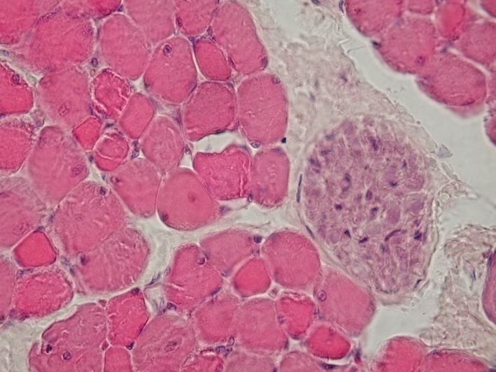
SKELETAL MUSCLE
SMOOTH MUSCLE
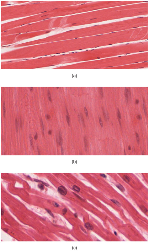
SKELETAL, SMOOTH AND CARDIAC MUSCLE
The Neuromuscular Junction: Pp 140-141 in the lab manual
- Somatic motor neuron - stimulates each muscle fiber to contract
- Motor unit - a motor neuron PLUS all the skeletal muscle fibers it innervates
- Axon terminals - the axon of a motor neuron divides into many branches within the muscle, each of which forms a NMJ
- NMJ (neuromuscular junction) - along with a skeletal muscle fiber, includes the axon terminals (branches) and the muscle fibers they innervate, and here each axon terminal divides into synaptic end bulbs (swellings filled with neurotransmitters in synaptic vesicles)
- Synaptic end bulbs - swellings at the end of axon terminals, filled with synaptic vesicles that are stimulated to release the neurotransmitters once the nerve impulse reaches here
- Synaptic vesicles - storage units filled with neurotransmitters that are released and diffuse across the synaptic cleft to bind to receptors in the motor end plate
- Neurotransmitters - chemical signals
- Synaptic cleft - space or gap between the synaptic end bulb and the muscle fiber
- Motor end plate - receptors on the region of the sarcolemma (plasma membrane of the muscle fiber), where neurotransmitters bind
- Sarcolemma - plasma (cell) membrane on the muscle fiber
- Action potential - nerve impulse that is generated that stimulates skeletal muscle fiber to contract
- Contraction - an action potential that occurs simultaneously when a motor neuron stimulates all the skeletal muscle fibers in a motor unit
- Twitch contraction - a quick shortening and relaxation observed in a skeletal muscle when a single action potential traveling down a motor neuron stimulates the skeletal muscle fibers of the motor unit to contract. Three phases:
- Latent period - 2 milliseconds (2 msec) and is the time between stimulation of a muscle cell and force generation
- Contraction period - 10-100 msec; the period during which force (measured in grams) is increasing
- Relaxation period - 10-100 msec; the period when force is decreasing
- Wave summation - muscle fibers of a motor unit are stimulated before this phase is completed, so the next contraction produces a greater force, increasing the frequency of muscle stimulation, producing sustained force generation
- Unfused tetanus - there is a partial relaxation between muscle twitches; MOST sustained voluntary skeletal muscle contractions are of this type with different motor units stimulated at different times (asynchronous contractions), delaying muscle fatigue (inability to contract caused by long periods of muscle contraction)
- Fused tetanus - there is no relaxation observed between twitches
- Normal muscle contraction - not twitch contractions; sustained contractions of varying force
- Motor unit recruitment - increases the number of motor units contracting at the same time, which increases the amount of force generated
- Maximal force - occurs when ALL motor units of a muscle are stimulated and ALL muscle fibers are contracting
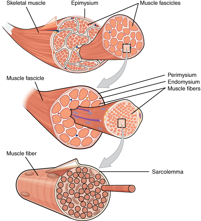
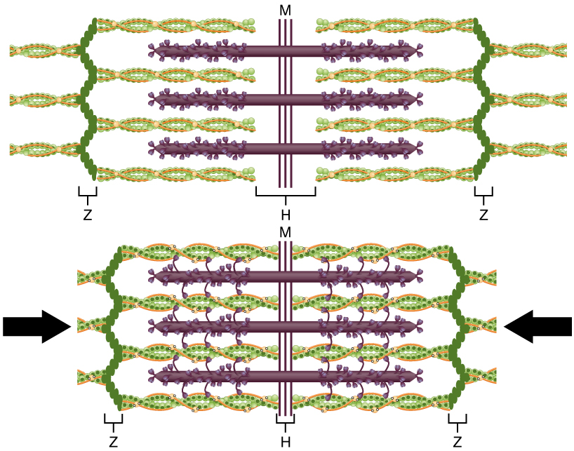
Connective Tissue Coverings of Skeletal Muscle:
Objectives:
FACIAL:
- Each skeletal muscle is considered to be an organ since it is composed of muscle tissue, connective tissue, nerve fibers and blood vessels.
- When stimulated by somatic motor neurons to contract, they produce a variety of motions or will stabilize body positions.
- Electrical impulses that initiate a contraction in one skeletal muscle fiber are not spread to an adjacent muscle fiber, due to 3 layers of connective tissue that separate and electrically insulate skeletal muscles:
- Endomysium
- Endo = within or inside
- Myso/myo = muscle
- Inner connective tissue that covers each individual skeletal muscle fiber
- Fascicles = bundles of skeletal muscle fibers surrounded by a layer called the perimysium
- Perimysium
- Peri = around
- Middle layer surrounding the bundles of skeletal muscle fibers called fascicles
- Epimysium
- Epi = upon; on; surrounding
- Connective tissue layer surrounding a whole muscle
- Formed by a number of fascicles
- Endomysium
Objectives:
- Describe how skeletal muscles are named
- Identify major skeletal muscles on models and charts
- Describe the actions of the major skeletal muscles
- Named for their orientation relative to the midline of the body
- Size
- Shape
- Action
- Facial expression/emotions
- Mastication (chewing)
- Flexion and extension and hyperflexion of the head and neck
- See Table 13.2 for Muscles of the Head and Neck
- Number of origins
- Location
- Superficial (shallow)
- Deep
- Origin AND insertion, usually skin or bone
- Origin - the nonmoving point of attachment when a muscle contracts
- Insertion - the point that moves toward the origin
- See the Table 13.1 on Page 148
FACIAL:
- Frontalis - in front; raises the eyebrows and wrinkles the forehead; lies over the frontal bone
- Occipitalis - in back; lies over the occipital bone; pulls scalp posteriorly
- Orbicularis oculi - encircles the eye; circular muscle; closes the eye and eyelid
- Zygomaticus - between zygomatic bone and corner of the mouth; raises corners of the mouth; "smiling muscle"
- Orbicularis oris - encircles the mouth; circular muscle; closes and purses the lips; "kissing muscle"
- Temporalis - lies over the temporal bone; elevates and retracts mandible; aids in chewing; closes the mouth
- Masseter - between the zygomatic arch and posterior portion of mandible; elevates and retracts the mandible for chewing & closes the mouth
- Sternocleidomastoid - on anterior and lateral neck; fibers run diagonally across the neck between sternum, clavicle, mastoid process; strap-like muscle; both muscles contract and flex the head; "prayer muscle"; one contracting rotates the head side-to-side as in saying "no"
- Trapezius - superior portion; superficial muscle; triangular-shaped muscle that is part of the upper back and posterior neck that extends the head
TRUNK MUSCLES:
- Muscles that move the arm at the shoulder joint
- Deltoid - anterior; flexes arm; lateral portion abducts arm; posterior portion extends arm
- Pectoralis major and minor - chest; adducts and flexes arm at the shoulder joint
- Latissimus dorsi - extends and adducts the arm at the shoulder joint or if elevated over the head, brings it down; posterior
- Muscles that move the scapula
- Serratus anterior - "saw-toothed" muscle like a "serrated knife"; on lateral trunk inferior to arms and ribs; abducts scapula and rotates it upward; "boxer's muscle"
- Trapezius - posteriorly, this diamond-shape muscle of the posterior neck and upper back extends from the skull to the spine of the scapula to the vertebral column and the superior portion elevates the scapula, middle portion adducts it, inferior portion depresses it
- Muscles that move the abdominal wall and vertebral column
- External oblique - lateral and anterior sheet-like abdominal muscles whose fibers run obliquely toward the midline; both contract and flex the vertebral column and compress the abdomen; one contracting laterally flexes the vertebral column
- Rectus abdominis - midline abdominal muscles located between the sternum and the groin; "six pack"; flexes vertebral column and compresses abdomen; does not laterally flex the vertebral column
- Internal oblique - deep; abdominal muscles run deep to the external oblique, whose fibers run obliquely toward the midline; both muscles contracting flex vertebral column and compress abdomen; one muscle contracting laterally flexes vertebral column
- Transversus abdominis - deep; abdominal muscles deep to the internal oblique whose fibers run transversely; compresses abdomen only
- Erector spinae - posteriorly, it is a group of deep muscles next to the vertebral column and deep to the trapezius, latissimus dorsi, and scapula that extend the vertebral column and maintain erect posture when both muscles contract; laterally flexes the vertebral column when only one muscle contracts
Muscles of the Arm:
Anterior Surface:
Posterior Surface:
Muscles of the Forearm:
Anterior:
Posterior:
Medial:
Anterior and Lateral Superficial Muscles:
Anterior Surface:
- Biceps brachii - flexes the forearm at the elbow and flexes the arm at the shoulder joint; largest muscle in the anterior compartment of the arm; has 2 heads
- Biceps = two heads
- Brachii = arm
- Brachialis - flexes the forearm at the elbow; posterior to the biceps brachii and anterior to the humerus
Posterior Surface:
- Triceps brachii - extends the forearm at the elbow and extends the arm at the shoulder joint; largest muscle with three heads
- Triceps = three heads
Muscles of the Forearm:
- Brachioradialis - a large muscle found in the forearm that flexes it; superficial lateral muscle
- Brachi = arm
- Radialis = radius
- Flexors of forearm: flexor carpi, radialis, carpi ulnaris, palmaris longus
- Carpi = wrist
- Flexes the hand at the wrist joint
- Extensors of forearm: extensor carpi ulnaris, extensor digitorum, extensor carpi radialis longus, extensor carpi radialis brevis
- Extends the hand at the wrist joint
Anterior:
- Quadriceps femoris group: 4 muscles of the anterior thigh
- Quad = 4
- Cep or caput = head or origins
- Rectus femoris - located along the midline of the thigh; extends leg at knee and flexes thigh at hip
- Vastus lateralis - lateral to rectus femoris; extends leg at knee only
- Vastus medialis - medial to rectus femoris; extends leg at knee only
- Vastus intermedius - deep to rectus femoris; intermediate to vastus lateralis and vastus medialis; extends leg at knee only
- Tensor fasciae latae - small lateral hip muscle that flexes and abducts the thigh at the hip joint
- Tensor = to stretch
- Fascia = band
- Latus = wide
- Sartorius - diagonal muscle running from tensor fascia latae to beyond the medial knee; flexes leg at knee, abducts, and laterally rotates thigh at hip; allows us to flex and cross our legs
- Sartor = tailor
- Adductors -group of muscles on the medial surface of the thigh that include the pectineus, adductor magnus, adductor longus, adductor brevis, and gracilis that adduct the thigh at the hip
Posterior:
- Hamstring muscle group - 3 muscles of the posterior thigh
- Biceps femoris - most lateral hamstring; flexes leg at knee and extends thigh at the hip
- Semitendinosus - medial to the biceps femoris and superficial to the semimembranosus; flexes leg at knee and extends thigh at hip
- Semimembranous - medial to biceps femoris and deep to semitendinosus; flexes leg at knee and extends thigh at hip
- Gluteus maximus - largest buttocks muscle; extends thigh at hip joint only
- Gluteus = buttocks
Medial:
- Adductor magnus
- Adductor longus
- Adductor brevis
- Pectineus
- Gracilis
Anterior and Lateral Superficial Muscles:
- Tibialis anterior - Lateral to the anterior crest of the tibia; dorsiflexes and inverts the foot
- Extensor digitorum longus - lateral to the tibialis anterior; dorsiflexes the foot and extends the toes
- Fibularis (peroneus) longus - Lateral to the extensor digitorum longus; plantar flexes and everts the foot
- Gastrocneumius - large superficial muscle on posterior leg; plantar flexes the foot and flexes the leg at the knee
- Soleus - deep to the gastrocnemius and posterior to the fibularis (peroneus) longus; plantar flexes foot only
- Calcaneal (Achilles) tendon - tendon of the gastrocnemius and soleus muscles
