Biology: Themes in the Study of Life
PHYSIOLOGY: THE CHEMISTRY OF LIFE
Biology is the study of life and of living organisms. It includes the study of the structure and function of a living organism, growth, evolution, distribution, ecology, and taxonomy. It is vast and there are a number of disciplines and sub-disciplines. Physiology is the study of the normal function of living organisms and their metabolic activities. It is a sub-discipline of biology. It is a focus of how cells, tissues, organs, organ systems, and the living organism carry out their normal chemical and physiological functions.
THE CELL: The Basic Unit of Life (Examples of Cells)

By Zaldua I., Equisoain J.J., Zabalza A., Gonzalez E.M., Marzo A., Public University of Navarre - Own work, CC BY-SA 4.0, https://commons.wikimedia.org/w/index.php?curid=46386894
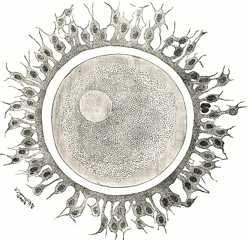
Ovum: Largest Cell in the Human Body
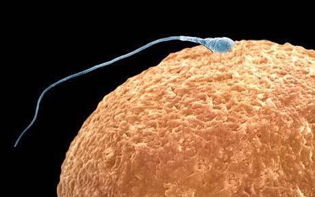
Sperm: The only flagellated cell in the human body (male)
|
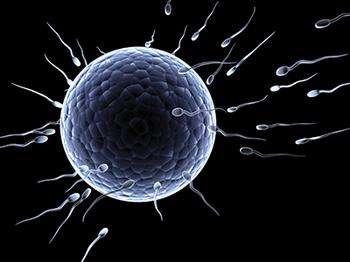
Sperm: The only flagellated cell in the human body (male)
|
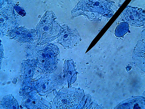
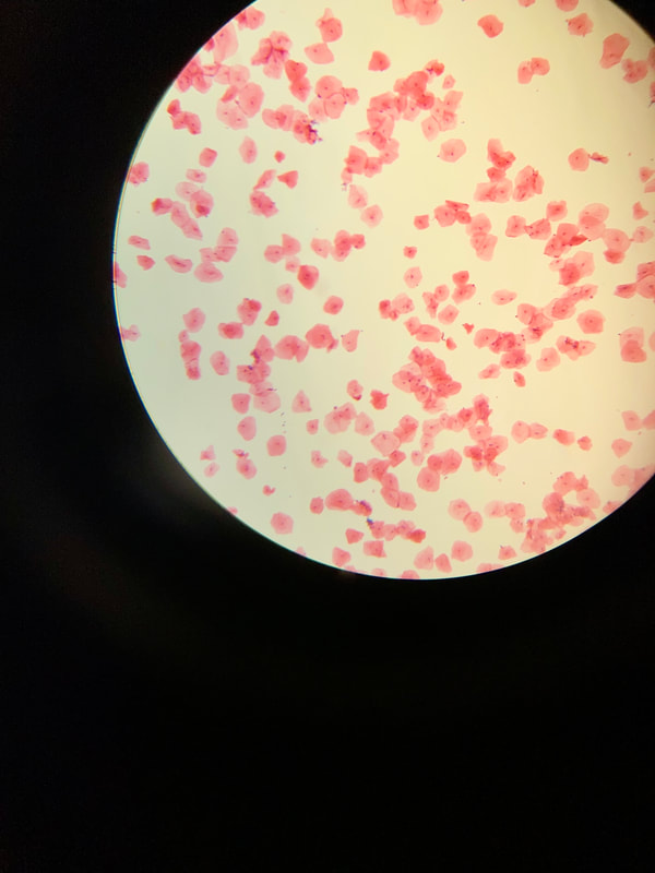
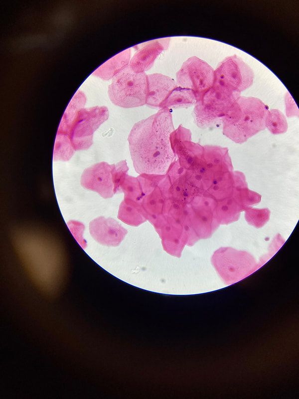
Squamous Epithelial Cells
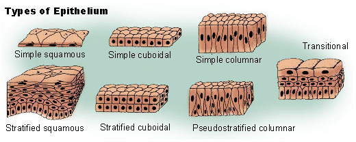
Epithelium
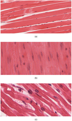
a) Skeletal muscle cells; b) Smooth muscle cells; c) Cardiac muscle cells
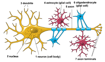
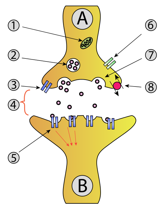
Neurons
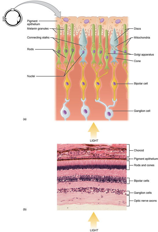
Rods and Cones in the Eyes
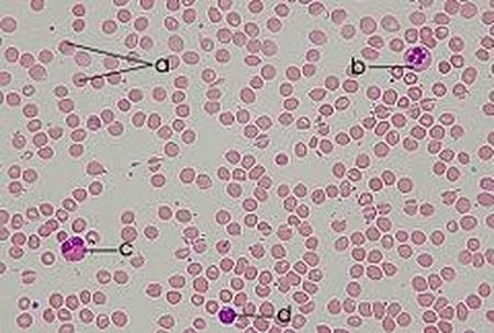
a) Red blood cells; b) White blood cell: Neutrophil; c) White blood cell: Eosinophil; d) White blood cell: Basophil; Small purple circles: Platelets

Plant Cell
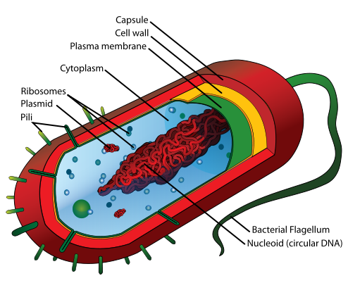
Prokaryote Cell (Bacteria)
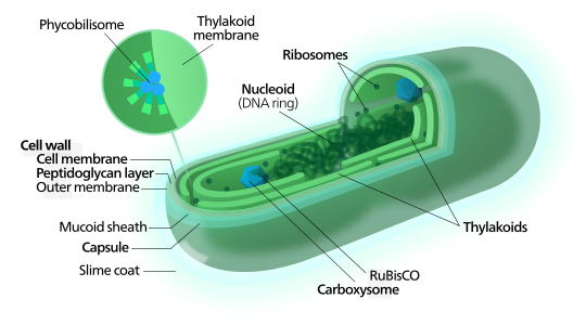
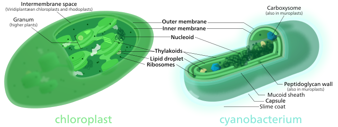
Cyanobacteria
Eukaryote
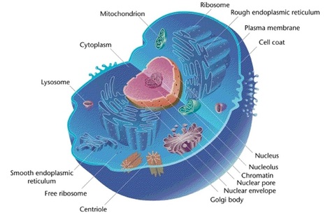
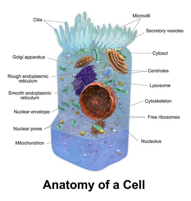
THE CELL: The Basic Unit of Life
By Zaldua I., Equisoain J.J., Zabalza A., Gonzalez E.M., Marzo A., Public University of Navarre - Own work, CC BY-SA 4.0, https://commons.wikimedia.org/w/index.php?curid=46386894
|
|
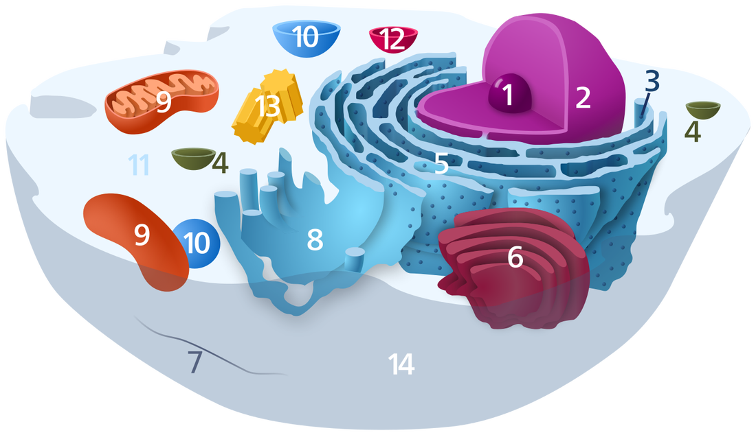
- 1) Nucleolus (RNA)
- 2) Nucleus (DNA)
- 3) Ribosomes, Associated With Rough Endoplasmic Reticulum
- 4) Vesicle
- 5) Rough Endoplasmic Reticulum
- 6) Golgi Apparatus
- 7) Plasma Membrane or Cell Membrane
- 8) Smooth Endoplasmic Reticulum
- 9) Mitochondria
- 10) Vacuole
- 11) Cytoplasm
- 12) Lysosome
- 13) Centrioles
- 14) Cell Wall
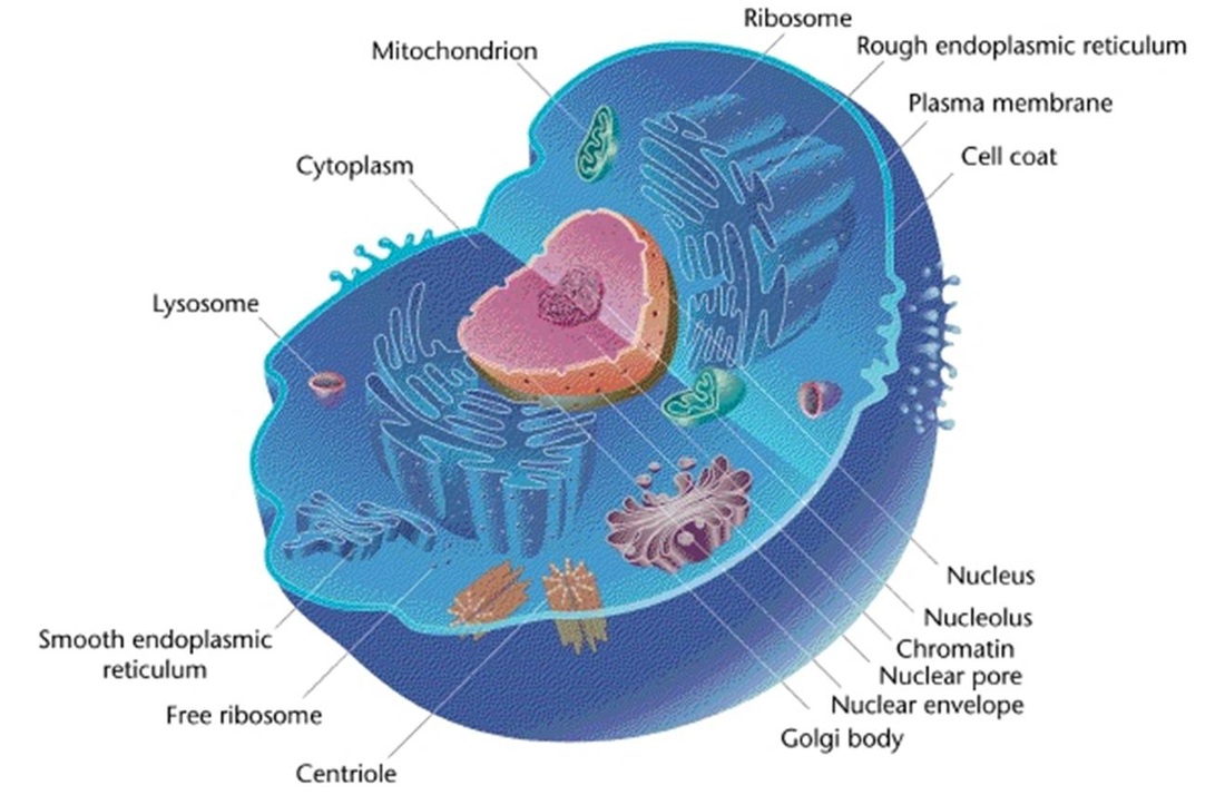
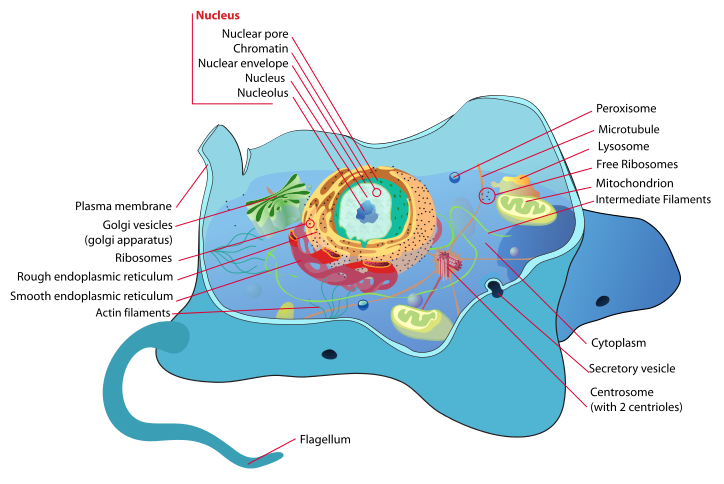
The cell is the basic structural and functional unit of the body and of every living organism. There are 2 types: prokaryotes and eukaryotes. Prokaryotic cells consist of bacteria and archaea, whereas eukaryotic cells consist of humans, animals, plants, protists, and fungi. Inside cells is a gel-like matrix composed of water and dissolved nutrients. It is a semifluid called the cytoplasm, or cytosol. In this jellylike substance, the subcellular components and cellular "organs" called organelles are suspended and perform their specialized functions. The term "eukaryote" means "true nucleus".
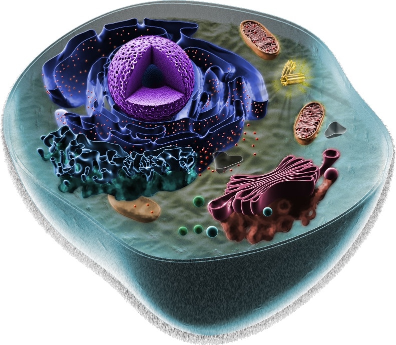
The Nucleus: The Command and Control Center of the Cell
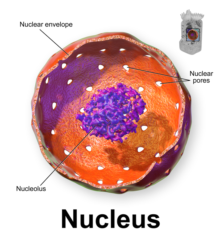
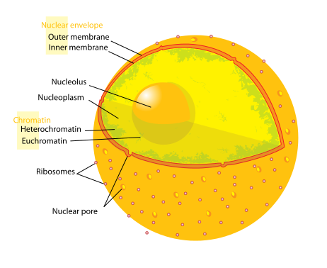
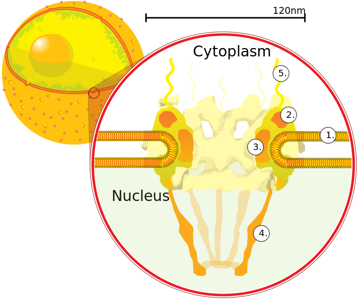
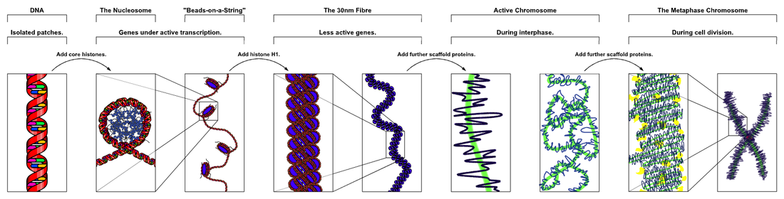
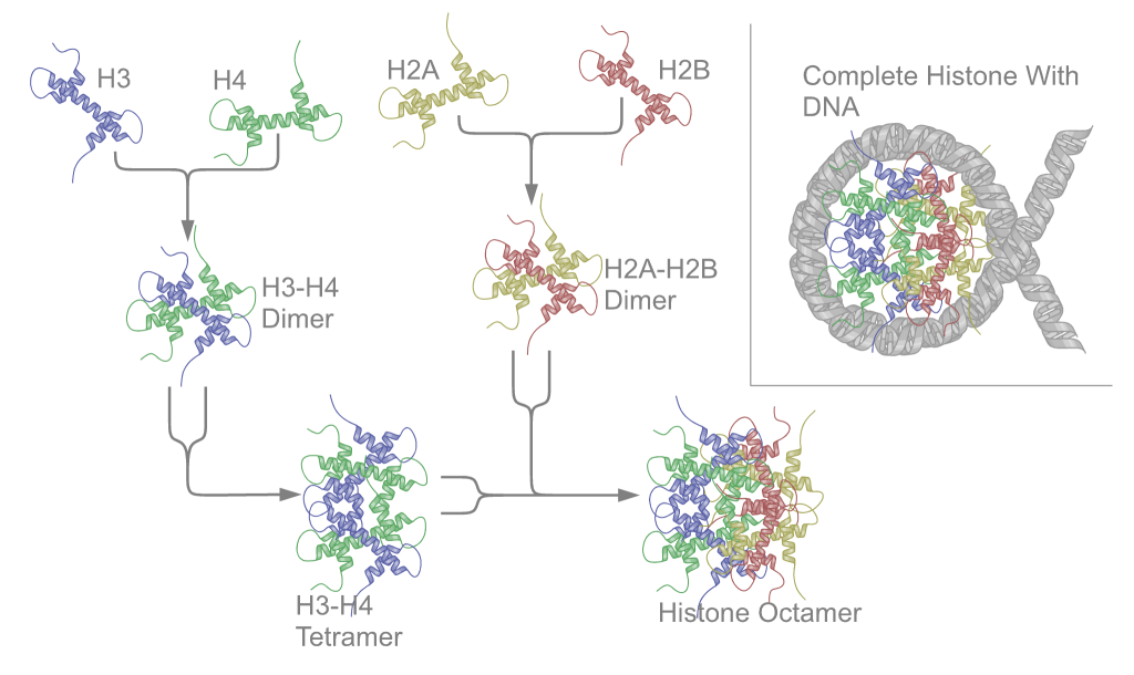
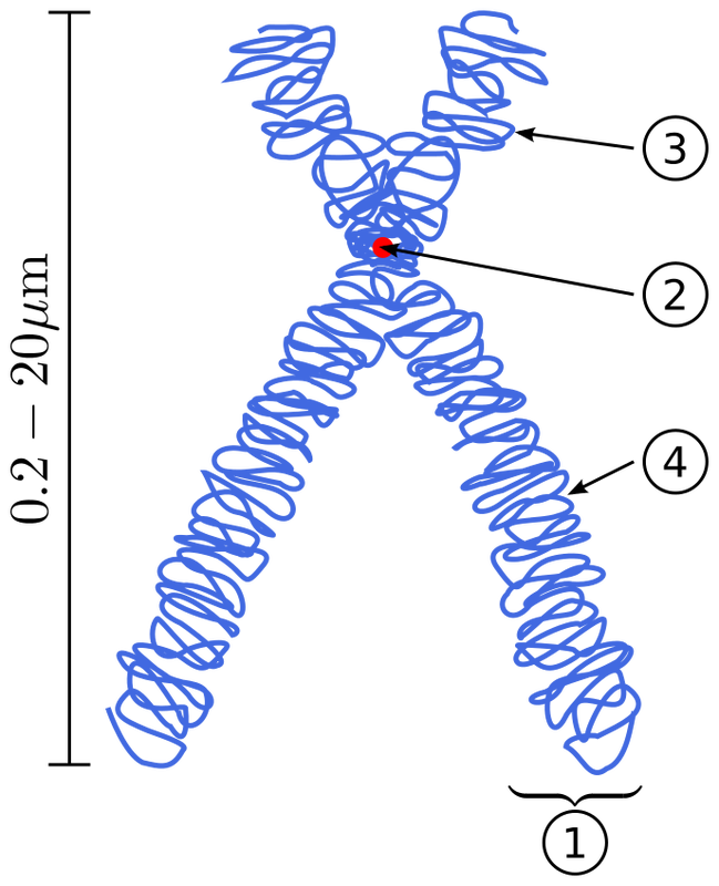
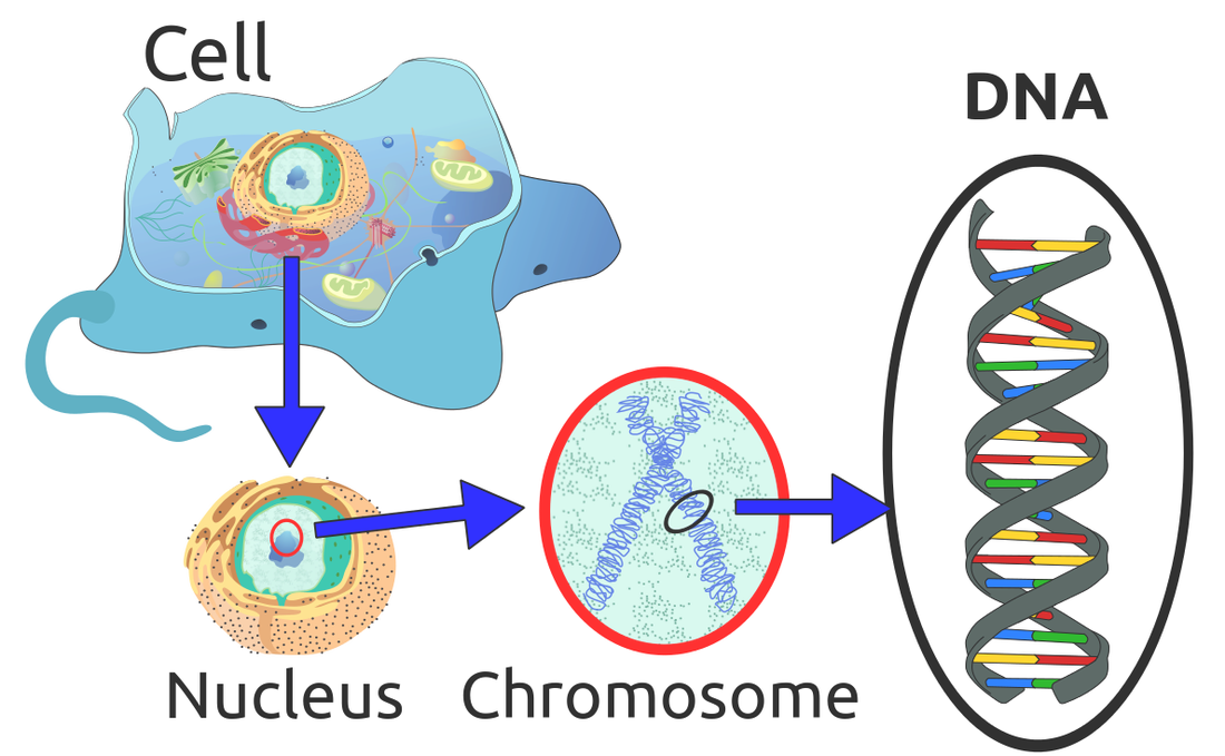
The nucleus is the command and control center of the cell. It consists of the porous nuclear envelope, the nucleolus core where RNA is housed, and chromatin, the material consisting of DNA and proteins, which is visible in dividing cells as individual condensed chromosomes. The nuclear membrane is a double-layered membrane surrounding the nucleus. It is perforated by basket-shaped pores and it connects to the endoplasmic reticulum. The nucleus is the area of the cell that contains most of the genes. The nuclear pores are lined with specialized pore complexes that line each pore and play a critical role in the regulation of proteins and RNA in and out of the nucleus. Inside the nucleus itself, the DNA is organized into units called chromosomes. Chromosomes carry the genetic information. Each chromosome consists of one long DNA molecule associated with many proteins, some of which are called histone proteins, which aid in the coiling of the DNA into chromosomes, which can fit into the nucleus.
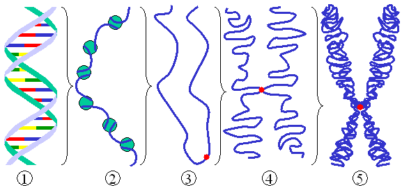
1) DNA, 2) Histone proteins for coiling of the DNA into chromosomes, 3) long DNA molecules/chromatin, 4) beginning to coil, 5) a chromosome with a centromere connecting in the middle
The Nucleolus:

The nucleolus is the heart of the nucleus. It is a non-membranous structure whose role is to produce ribosomes. A nucleus may contain one or multiple nucleoli. The ribosomal RNA (rRNA) is housed here. rRNA is synthesized here by the instructions provided by the DNA in the nucleus. Also here are proteins imported from the cytoplasm, which are assembled with rRNA into the large and small subunits of ribosomes.
Chromatin:
Chromatin is the material consisting of DNA and proteins that is found in the nucleus of the cell.
Chromosomes:
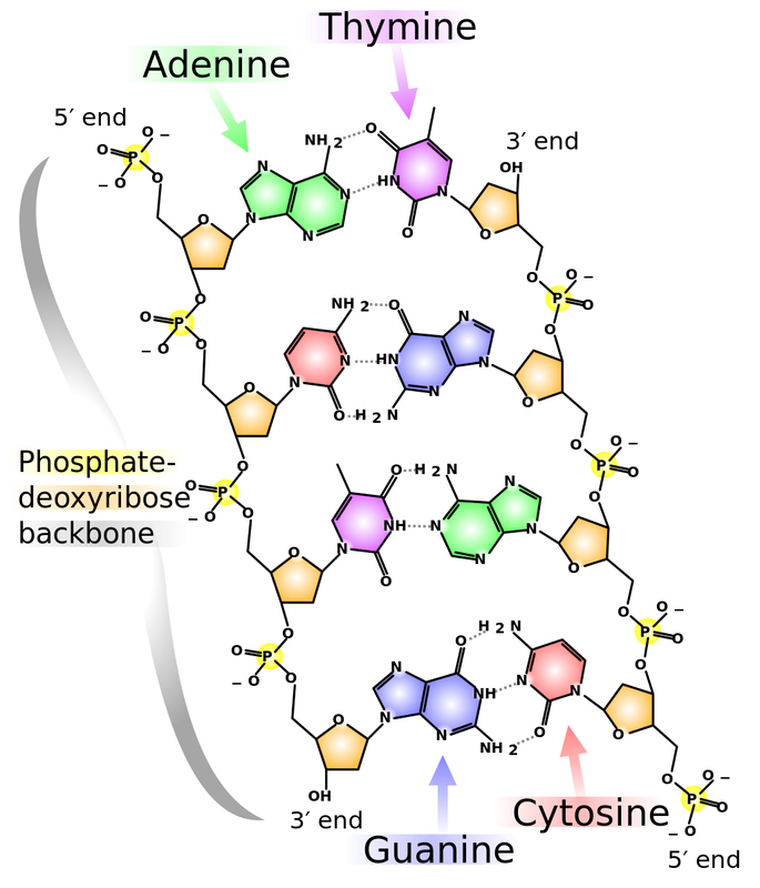
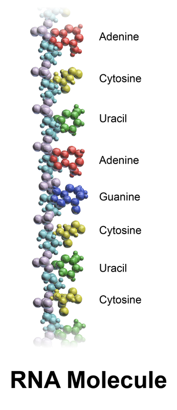
DNA and RNA:
|
|
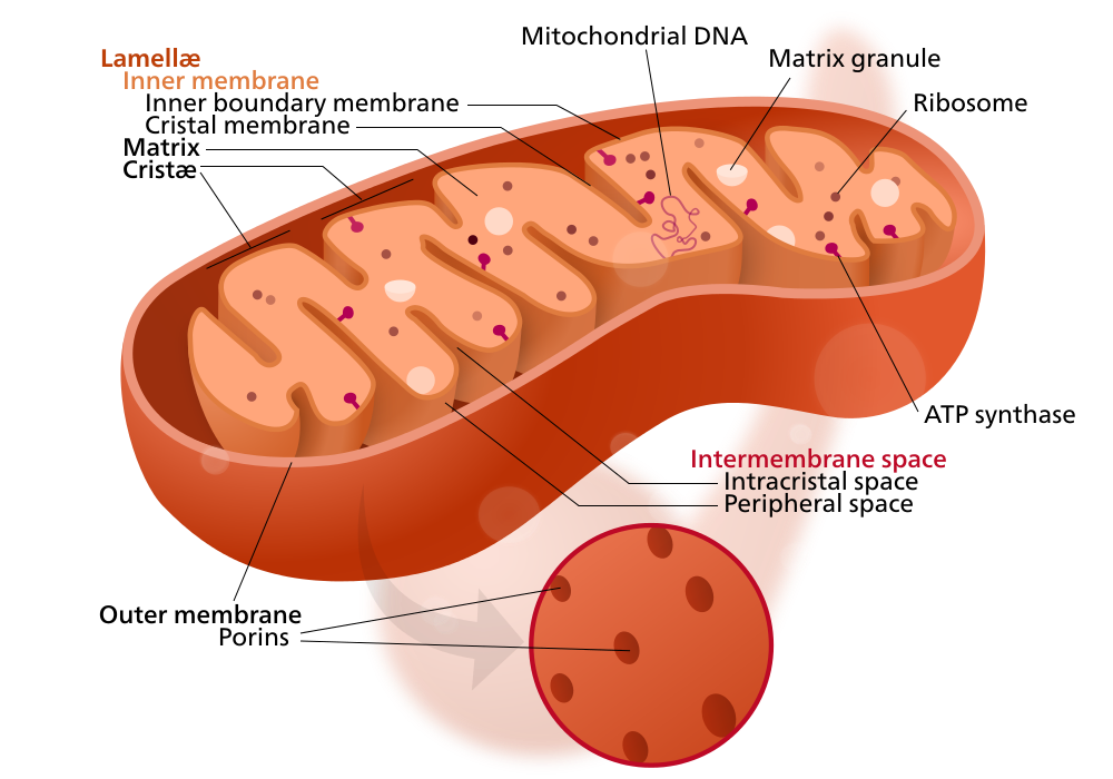
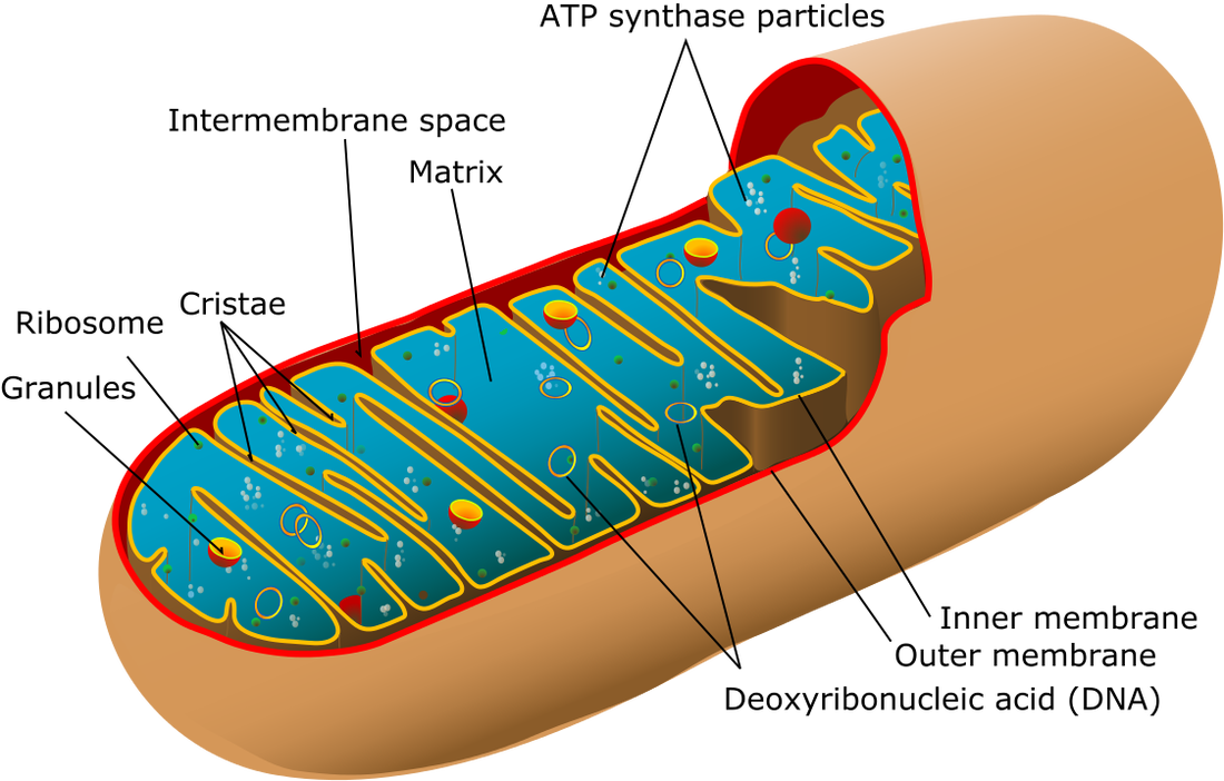
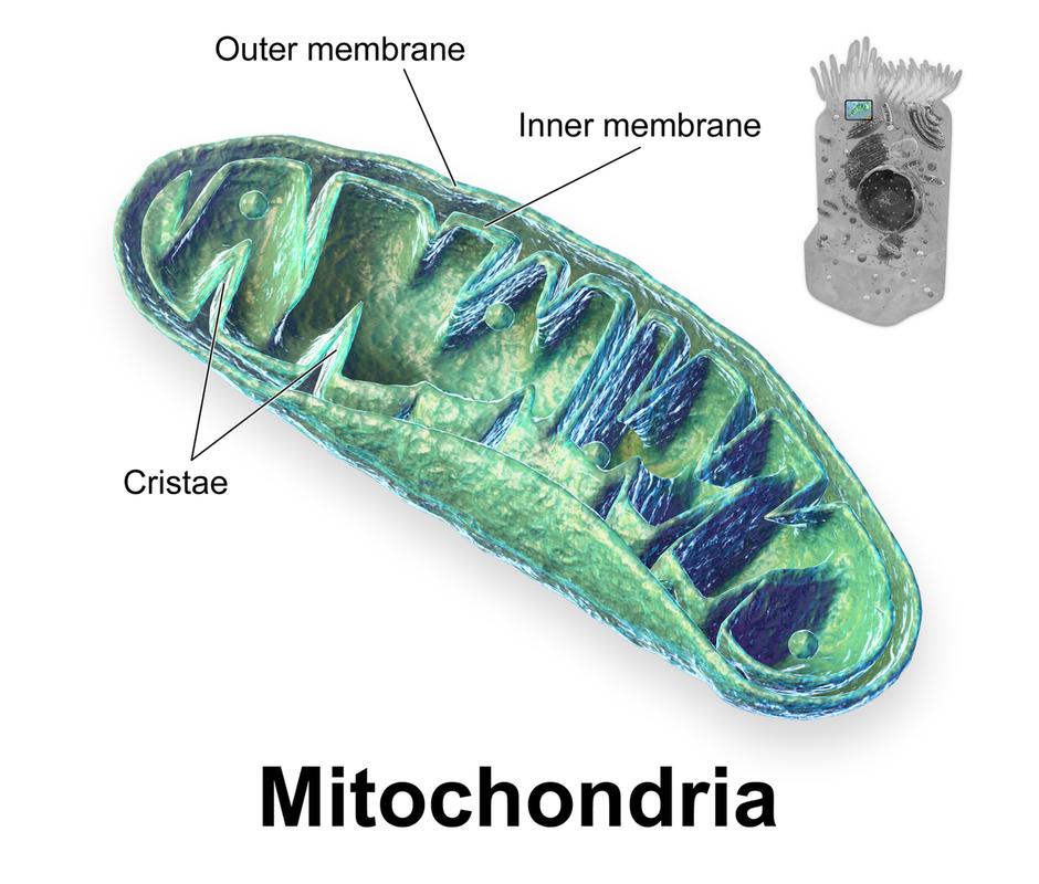
The Mitochondria: The Energy Powerhouse of the Cell
The mitochondria are the energy powerhouses of the cell. In this organelle, cellular respiration occurs and the most ATP energy is produced here in the cristae, or folds of the organelle.
- Sites of cellular respiration
- Convert energy to forms that cells can use for metabolic processes and for work
- Extracts energy from sugars, fats, fuels like iron
- A cell may contain 1 to thousands of mitochondria
- The number per cell depends upon the cell's level of metabolic activity (muscle cells have many more)
- Out membrane is smooth and inner membrane is convoluted with cristae
- Mitochondrial matrix is enclosed by the inner membrane and contains many enzymes that catalyze the steps of cellular respiration
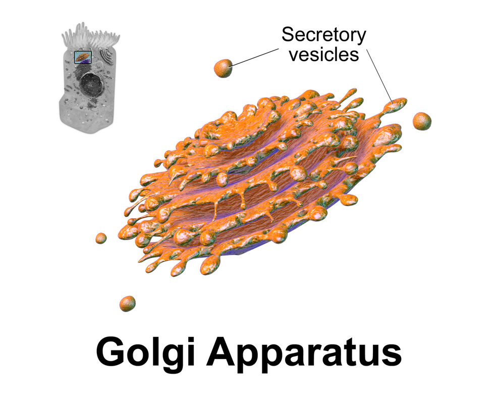
The Golgi Apparatus: The Packaging and Delivery Center of the Cell

|
|

|
|
|
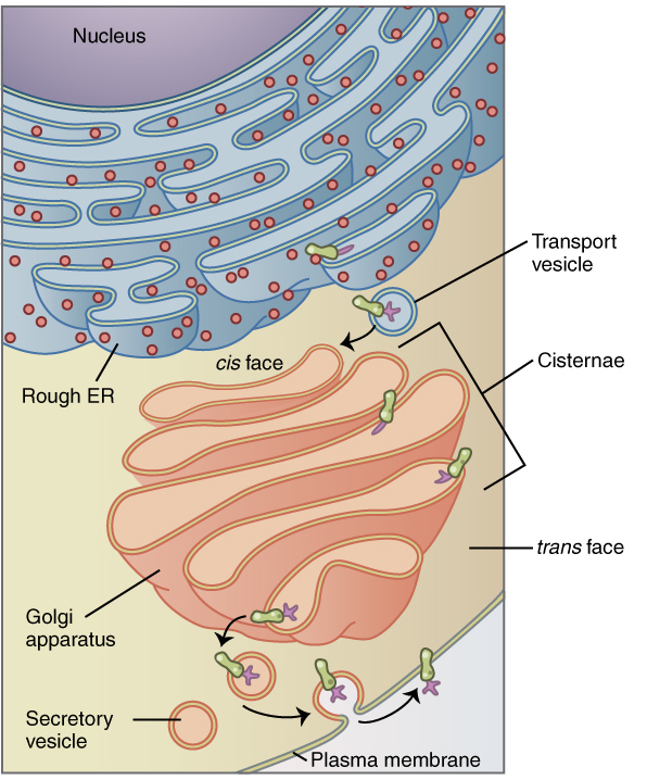
The Golgi Apparatus is like the UPS warehouse for receiving, packaging, sorting, manufacturing, shipping and the delivery system of the cell. Here, proteins are actively synthesized, modified, sorted, and secreted or delivered to other parts of the cell, or to the rest of the body. Also, some protein products from the ER are modified and stored, then sent to other destinations within or outside of the cell.
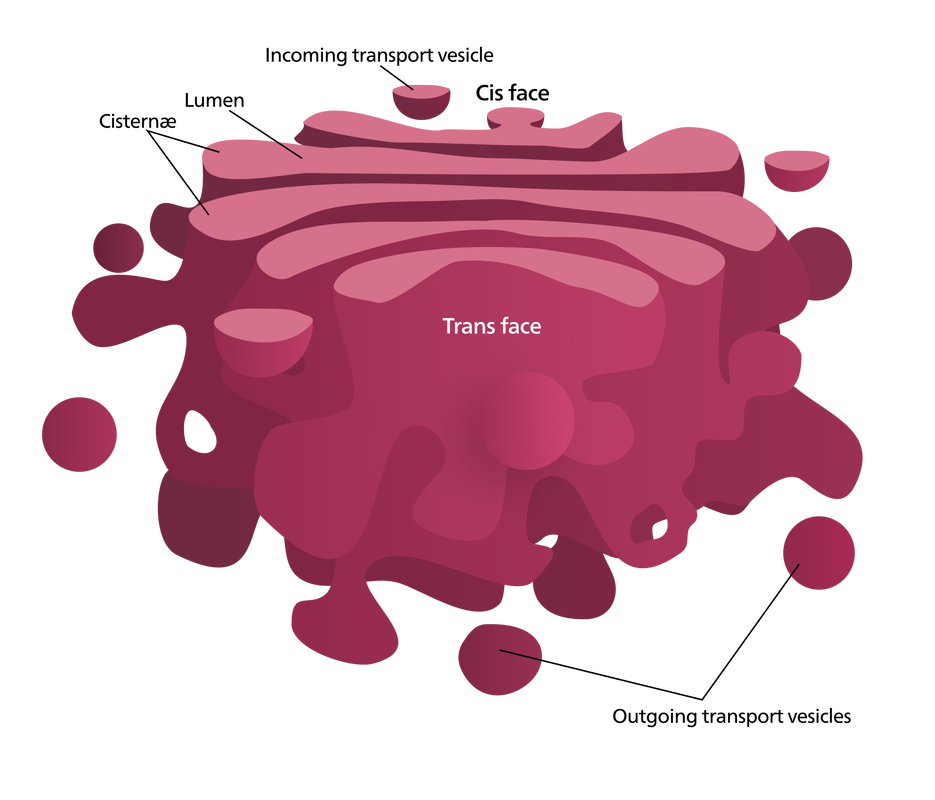
The Golgi Apparatus is a unique system of flattened membranous sacs (cisternae) that look like a stack of flat pita bread. The two sides of the stacks are referred to as cis face and trans face. The cis face is located near the ER and receives the proteins that need to be modified (endocytosis) and stored. The trans face breaks off into vesicles that pinch off (exocytosis) and travel to other areas within or outside of the cell.
Functions of the Golgi Apparatus:
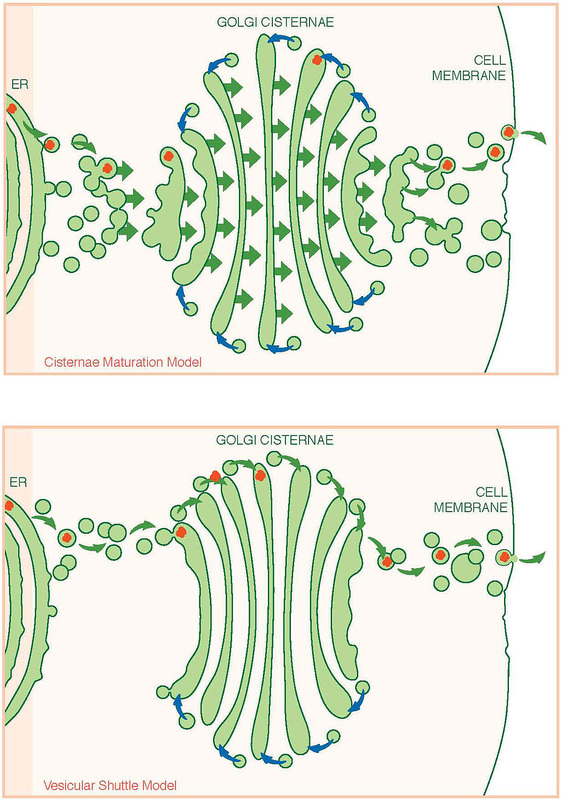
- Products of the ER are modified during their travel from the cis region to the trans region
- Glycoproteins formed in the ER have their carbohydrates modified here (some sugar monomers are removed, and some are substituted)
- Membrane phospholipids are altered here
- Manufacture of some macromolecules
- Polysaccharides may be synthesized and secreted from here
- Manufacturing and refining in stages
- Different cisternae contain different unique enzymes
- Protein products are sorted and targeted for various parts of the cell
- Molecular ID tags (phosphate groups) are added to Golgi products and aid in sorting
- Transport vehicles bud here with specific receptor and signaling molecules for transport
The Endoplasmic Reticulum: The Protein Alteration and Transport System of the Cell
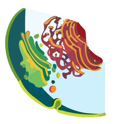
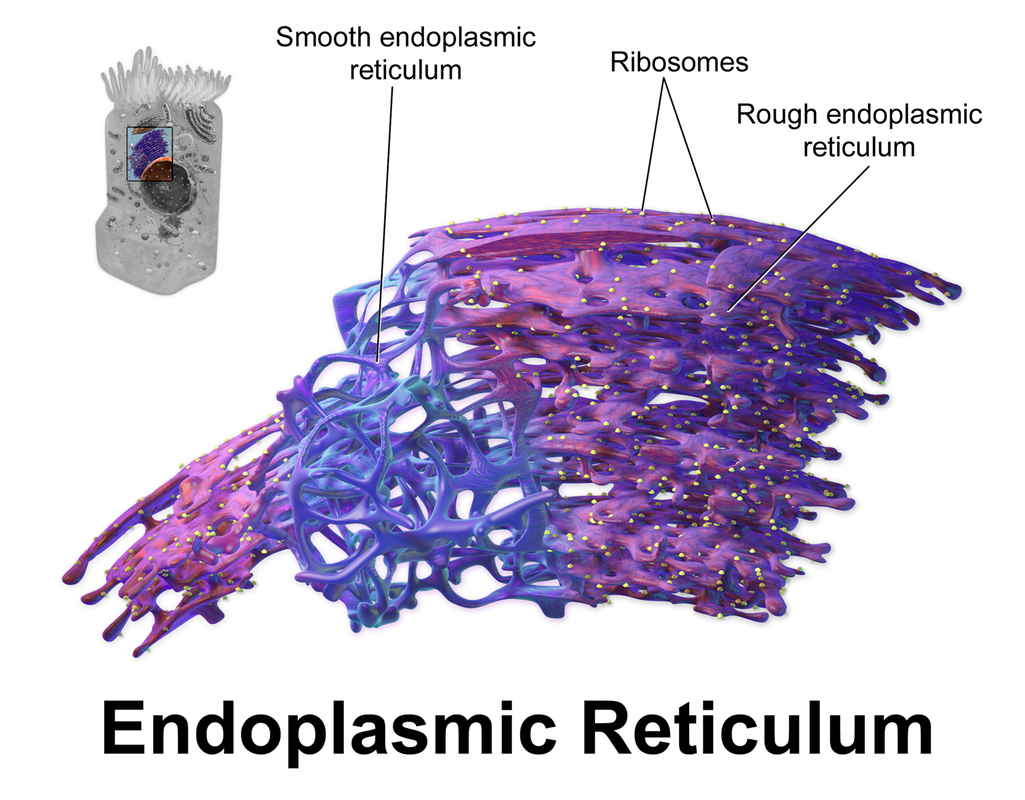
The endoplasmic reticulum is the transport system of the cell. Here, a network exists of membranous sacs and tubes that are active in protein synthesis and other synthetic and metabolic processes. There are 2 types: rough and smooth. The rough endoplasmic reticulum is studded with ribosomes, whereas the smooth endoplasmic reticulum has none. The endoplasmic reticulum system regulates protein traffic and performs metabolic functions in the cell, and it connects with the nucleus. The term "endoplasmic" means "within the cytoplasm" and the term "reticulum" is the Latin word for "little net". The membranous network of tubules and sacs are called "cisternae", from the Latin world "cisterna", meaning a "reservoir for liquid".
Functions of the Smooth ER:
Functions of the Rough ER:
Functions of the Smooth ER:
- Diverse metabolic processes
- Synthesis of lipids (fats), oils, phospholipids, steroids, cholesterol, sex hormones, steroid hormones secreted by the adrenal glands
- Metabolism of carbohydrates (sugars)
- Detoxification of drugs, alcohol and poisons, particularly in liver cells
- Storage of calcium ions in muscle cells
- Detoxification enzymes
Functions of the Rough ER:
- Protein synthesis (ribosomes)
- Insulin synthesis and secretion
- Polypeptide chain growth from ribosome is threaded through the lumen of the rough ER through a pore in the ER membrane, where it folds into its designated shape (4 types of protein folding)
- Protein secretion (glycoproteins are built here)
- Separation of secretory proteins from proteins produced by free ribosomes
- Production of vesicles, or bud-like bubbles that form from a specialized region called the transitional ER, where these vehicles for protein transport are formed
- Membrane factory for the cell: grows in place by adding proteins and phospholipids as needed
- Makes membrane phospholipids for the cell membrane
The Ribosomes: The Protein Synthesis Center of the Cell
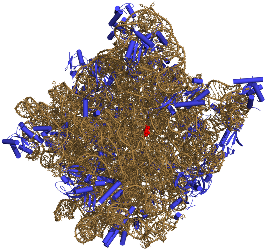
Ribosomes are small complexes that dot the rough endoplasmic reticulum, and they are active in the synthesis of proteins. They are also found free in the cytoplasm. Ribosomes are complexes made of ribosomal RNA (rRNA) and protein. They are the cellular organelles that carry out protein synthesis. Ribosomes bound to the rough ER tend to synthesize proteins that will be inserted into the cell membrane or those for secretion. Ribosomes contain 2 subunits: a large subunit and a small subunit.
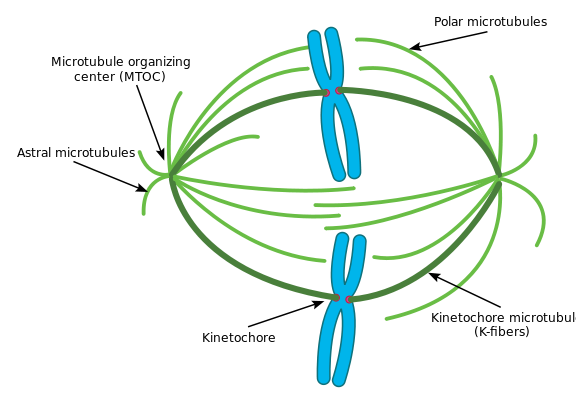
Centrioles:
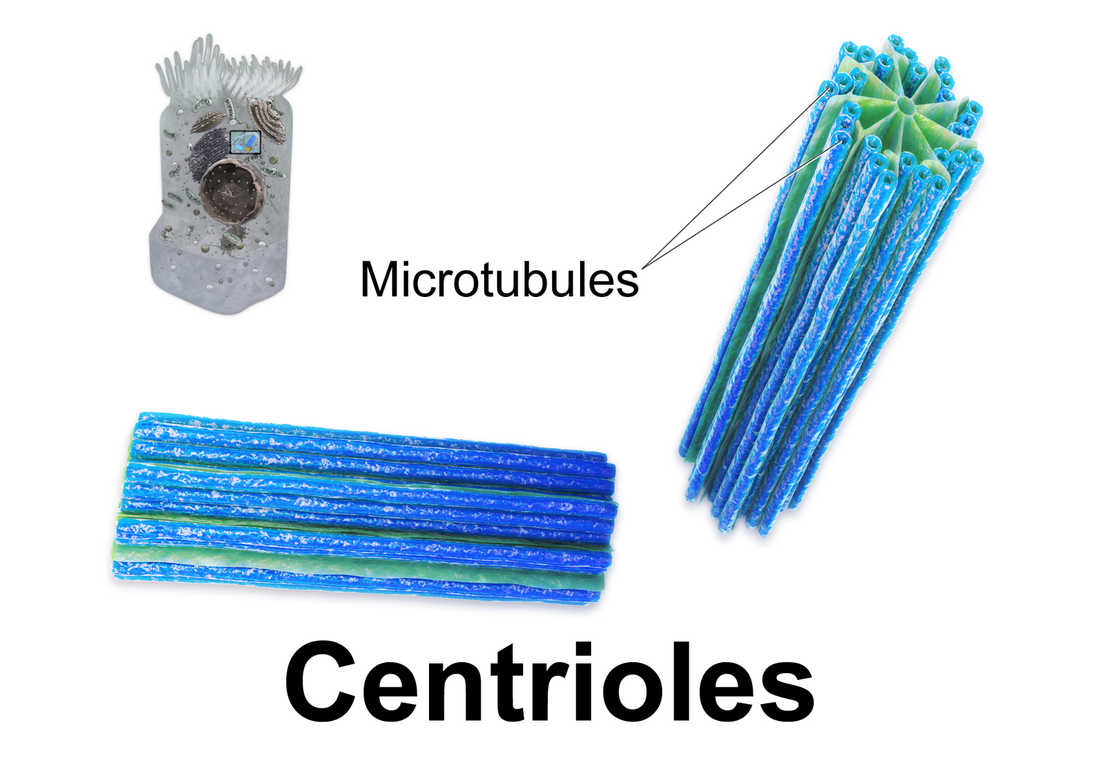
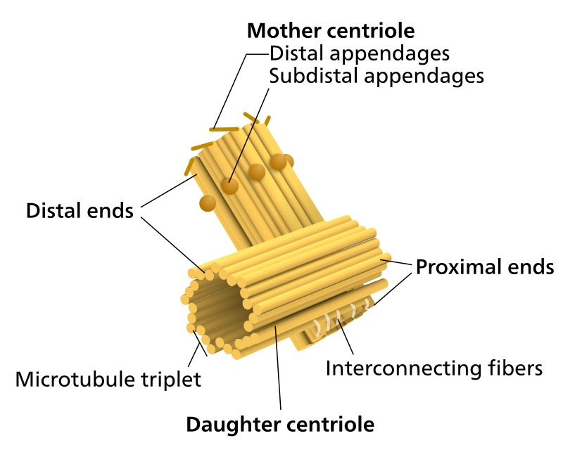
Centrioles or centrosomes are regions of the cell where the cell's microtubules are generated for cell division. They are in a pair, and they separate and go to opposite ends of the cell during this process.
- Found in the region near the nucleus
- "Microtubule-organizing center" of the cell
- Found in a pair
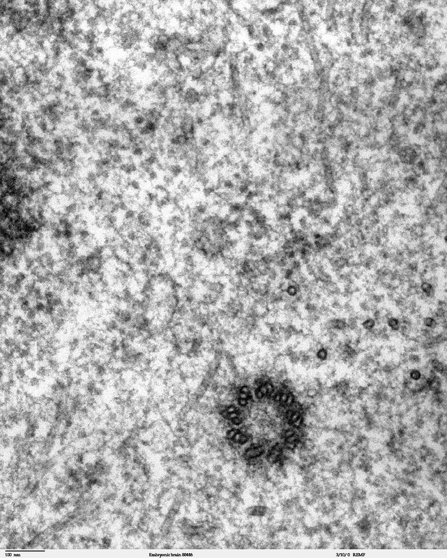
- Each is composed of NINE sets of triplet microtubules arranged in a ring
- Replicate prior to cell division
Microtubules:
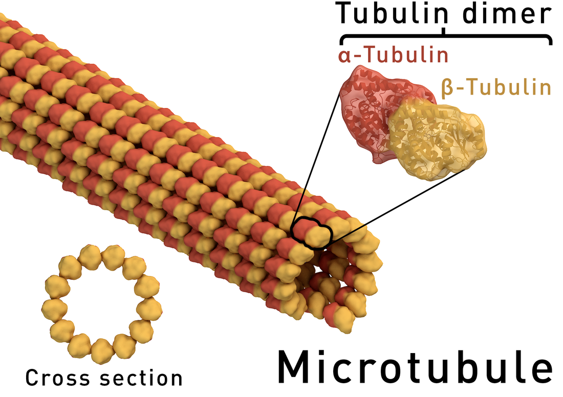
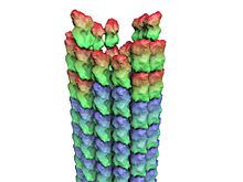
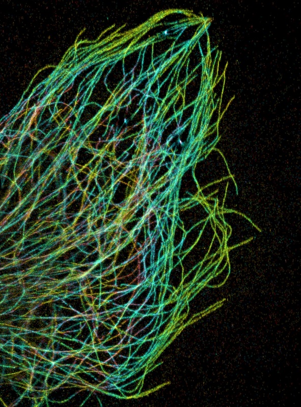
Microtubules are generated from the pair of centrioles in preparation for and during cell division. They also provide structure for the cell and are part of the cellular cytoskeleton, providing reinforcement for the cell's shape, function in cell movement, and its components are made of protein.
- They are the thickest of the 3 filament types in the cytoskeleton
- Hollow rods
- Constructed of the protein tubulin, each of which is a dimer (a protein made up of 2 subunits)
- Tubulin consists of a-tubulin and b-tubulin
- Microtubules grow in length by the addition of tubulin dimers
- Provide shape and support to the cell
- Serve as tracks in which motor proteins can move and be transported in the cell
- Guide secretory vesicles from the Golgi apparatus to the cell membrane for excretion
- Aid in separation of chromosomes during cell division
Microfilaments:
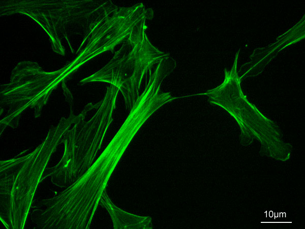
There are two types of filaments: microfilaments and intermediate filaments. They are made of protein and comprise the cell's cytoskeleton for reinforcement of shape, strength, motility, movement, and elasticity.
Microfilaments:
Intermediate Filaments:
Microfilaments:
- Thinnest filaments
- Actin filaments, a globular protein
- Role is to bear tension (pulling forces)
- Supports cellular shape
- Cell motility (muscle cells)
- Thicker filaments made of protein myosin
Intermediate Filaments:
- Filament fibers have diameters in a middle range
- Specialized for bearing tension
- Reinforce cell shape
- Members include the keratin proteins found in hair and nails
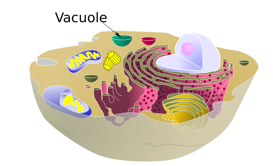
Vacuoles: Vesicles for Storage or Transport

Vacuoles are storage compartments in the cell for nutrients like iron, carbohydrates, and others, and for pigments, granules, enzymes, and things the cell might need later on for metabolic activities.
- Large vesicles that form from the pinching off of the ER and Golgi Apparatus
- Part of the cell's endomembrane system
- Selective in transporting solutes
- Food vacuoles (formed by phagocytosis)
- Contractile vacuoles (pump excess water out of the cell)
- Large central vacuole (plants) (cell sap and repository for inorganic ions such as potassium and chloride)
Lysosomes: Garbage, Breakdown, and Recycle Centers of the Cells

Lysosomes contain powerful, potent enzymes called lysozymes, which digest and breakdown cellular debris, foreign bodies, toxins and trash, and where macromolecules are hydrolyzed and destroyed.
Functions of the lysosome:
Functions of the lysosome:
- Membranous sac
- Hydrolytic enzymes
- Digest macromolecules
- Acidic environment
- Excessive leakage from a large number of lysosomes may result in autolysis, or self-digestion of a cell and cell destruction
- Intracellular digestion
- Phagocytosis and the resulting food vacuole fuse with a lysosome, whose enzymes then digest the food, such as simple sugars, amino acids, monomers, etc...
- Recycle the cell's own organic material (autophagy), resulting in the cell continually renewing itself
Peroxosomes: Alcohol Detox Centers of the Cell
Peroxisomes are specialized organelles containing potent enzymes that break down toxins such as alcohol, perform metabolic functions, and produce hydrogen peroxide as a by-product, then converting it to water.
- Some use oxygen to break fatty acids down into smaller molecules to be used as fuel for cellular respiration by mitochondria
- Peroxisomes in the liver detoxify alcohol, toxins and poisons by transferring hydrogen to oxygen
- Enzymes convert hydrogen peroxide to water
The Cell Membrane: The Security Perimeter of the Cell: Selective Permeability
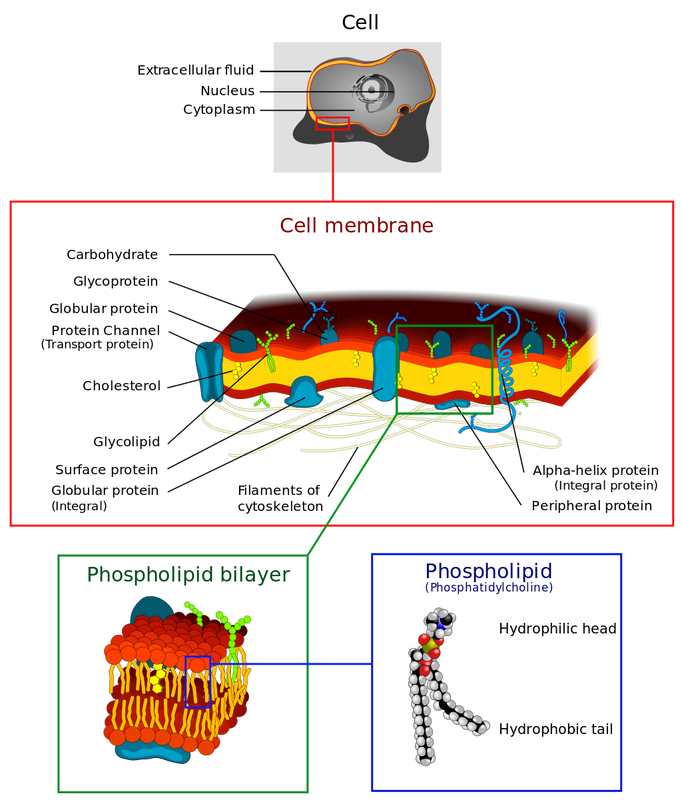
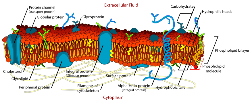
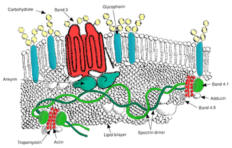
The cell membrane or plasma membrane is a secure perimeter surrounding the cell which protects it, helps it keep its shape, and is selectively permeable. It only allows certain things to come into the cell and allows toxins to exit the cell. Entrance and exit to and from the cell is highly monitored and regulated and occurs by specific cellular signals. The cell membrane is a double lipid membrane, containing proteins and receptors for signaling molecules, and cholesterol. The cell membrane functions as a selective barrier that allows passage of oxygen, nutrients and wastes to enter or exit the cell.
Vesicles:
Vesicles are transport vehicles of the cell. They transport proteins and hormones to other areas of the cell, into the cell, and out of the cell. Solid particles enter the cell via phagocytosis, or cellular "eating". This is endocytosis. Particles exit the cell via exocytosis. And liquid particles enter the cell via pinocytosis, or cellular "drinking". These are highly regulated processes and occur only through very specific signaling and receptor molecules present on the cellular membrane.
|

|
|

|
Flagella: Motility
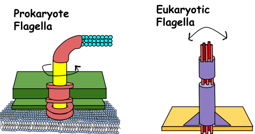
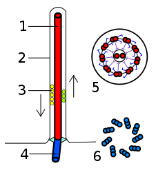
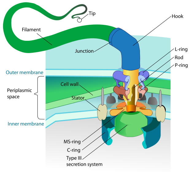
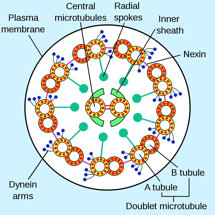
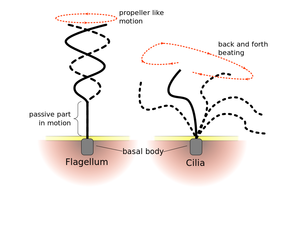
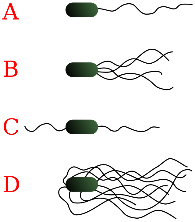
Flagella are motility structures present in some animal cells. They are composed of a cluster of microtubules that extend from the plasma or cell membrane.
- Specialized arrangement of microtubules is responsible for the beating movement of flagella and cilia
- Aid in the propelling of the cell through water or viscous substances
- Locomotor appendages
- Undulating motion like the tail of a fish that generates force or energy in the same direction as the flagellum's axis
Microvilli:
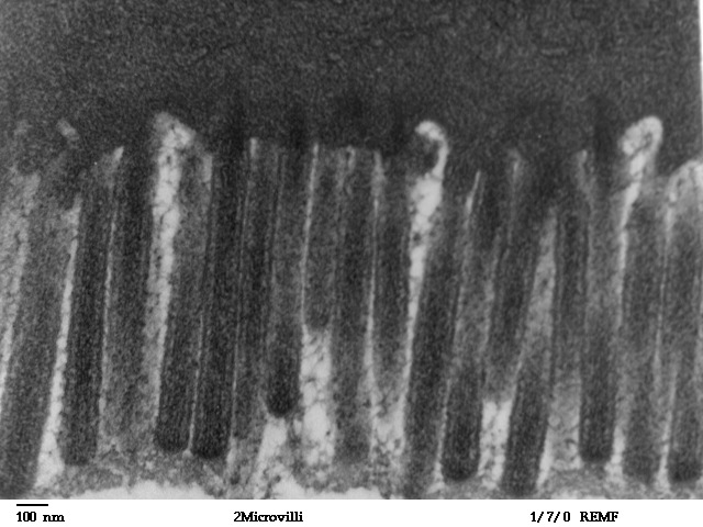
Microvilli are projections that extend from the cell membrane and they increase the cell's surface area for absorption of nutrients.
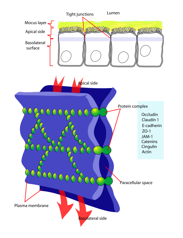
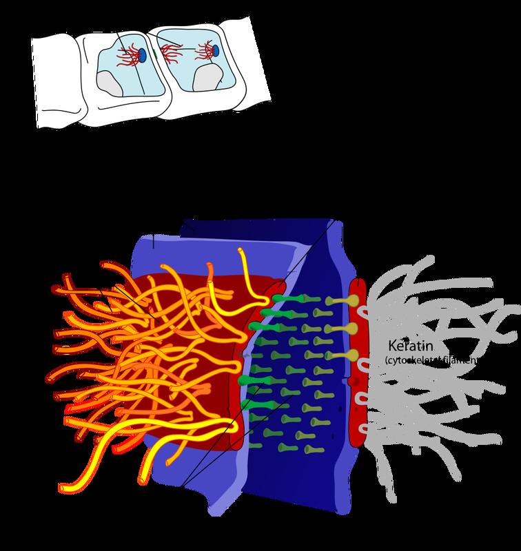
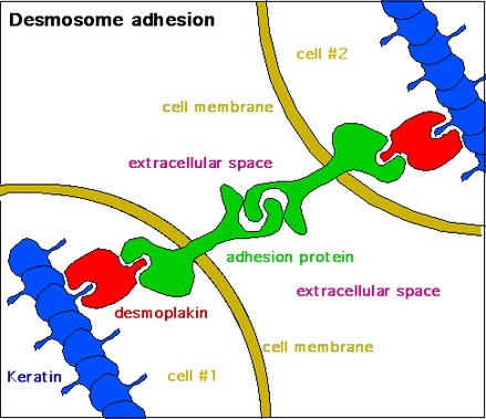
Cellular Junctions:
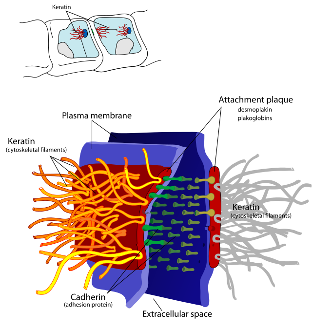

Desmosomes:
|
|
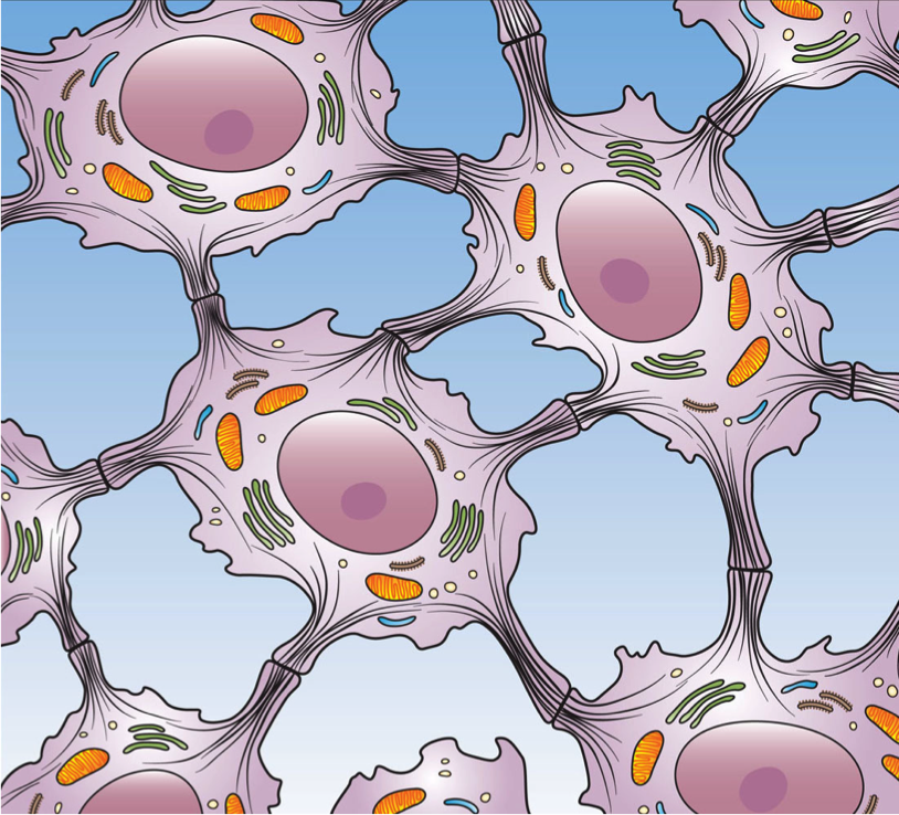
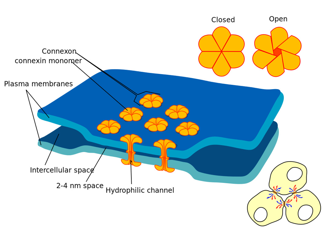
Gap Junction:
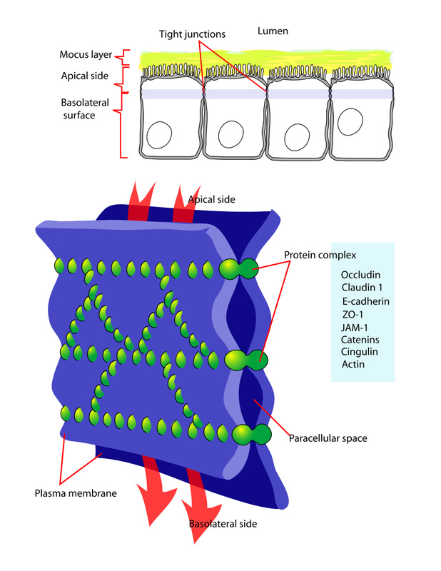
Tight Junction:
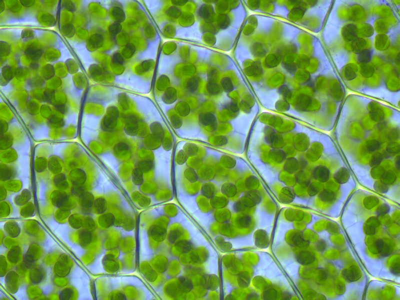
Plant Cell:
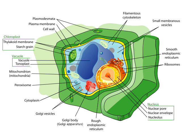
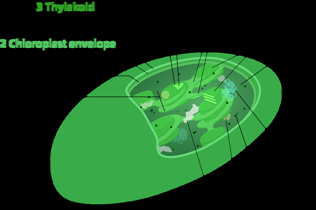
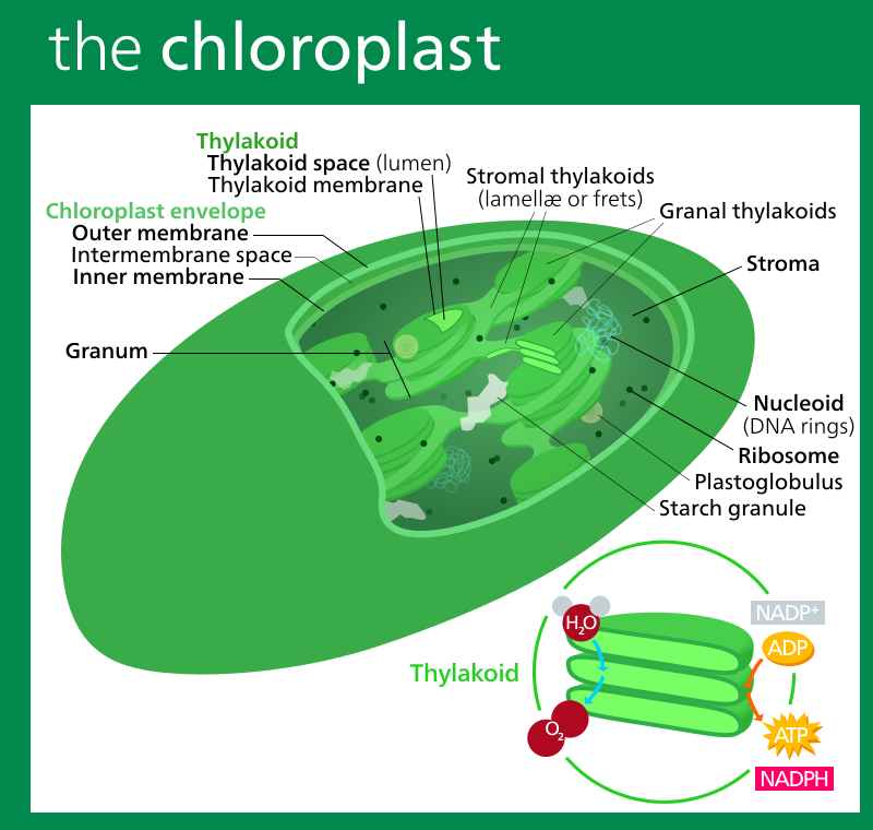
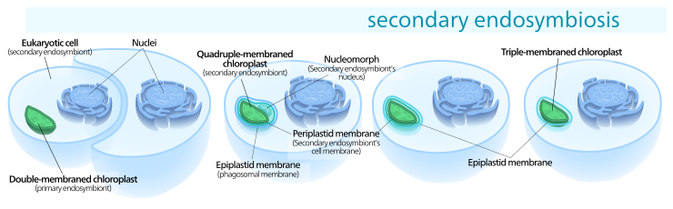
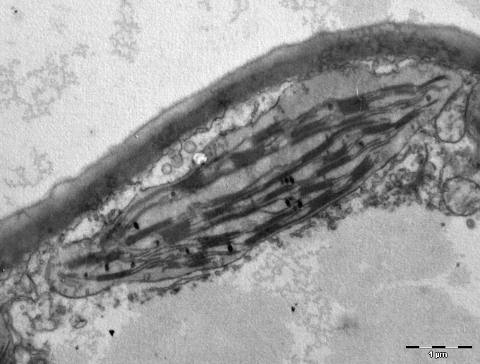
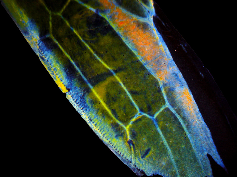
The Chloroplast:
|
|
|
|
|
|
The Thylakoid:
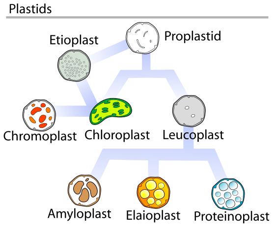
The Plastid:
|
|
Chitin: Cell Wall of Fungi
|
|
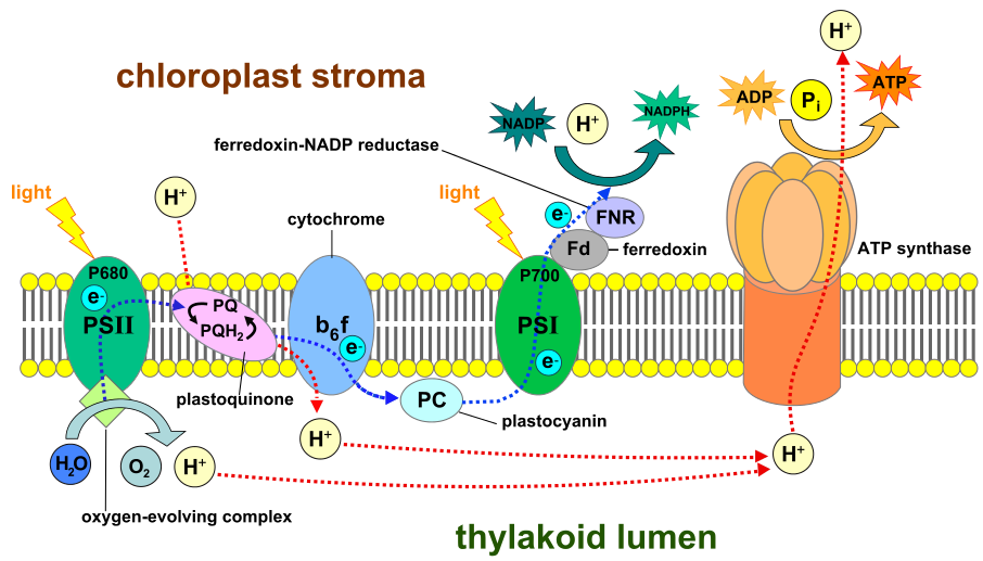
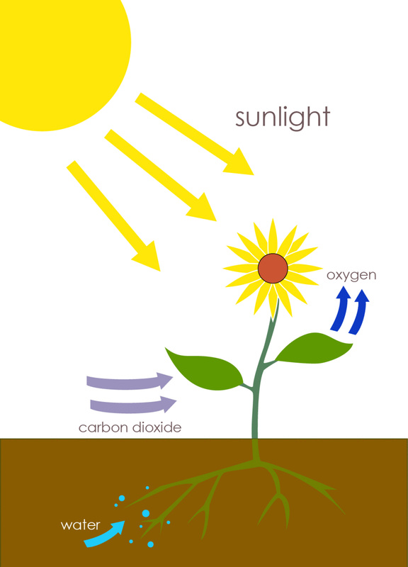
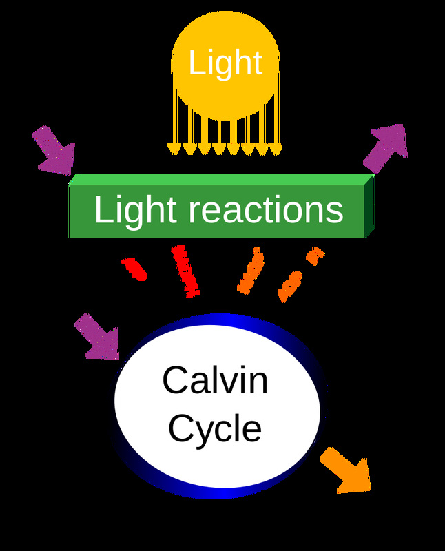
Photosynthesis:
|
|
Cell Transport:
The cell membrane is important because it is selectively permeable, meaning it is very "picky" about what it lets in and out of the cell. Nutrients are allowed to come in, but it keeps out things that might harm the cell, like toxins. Wastes are allowed to pass out of the cell. There are a variety of ways that things come into and exit the cell and are discussed below. Some require energy, whereas others do not.
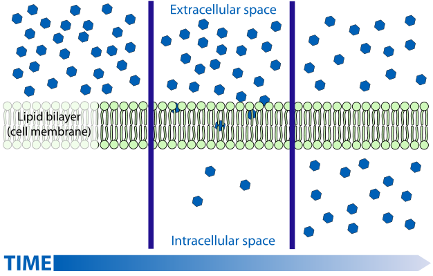
With simple diffusion, no energy is required for smaller molecules to move down the concentration gradient from an area of higher concentration to one of lower concentration, and some examples in the human body include oxygen (O2) and carbon dioxide (CO2). They diffuse across the cell membranes in the alveoli of the lungs and the red blood cells themselves as we inhale and exhale, and as the red blood cells pick up oxygen in the alveoli of the lungs and drop of CO2 to be exhaled. This is tied to the respiratory acidosis and alkalosis some of you wrote about in the last post during week 2, as interruptions of this process can cause a buildup of of carbon dioxide in the bloodstream, resulting in respiratory acidosis, or a loss of carbon dioxide in excess, resulting in respiratory alkalosis.
Another clinical picture of simple diffusion is the example of a negatively charged dye particle binding to the positive charges across the cell membrane and diffusing into it without any help of ATP energy or a protein carrier or channel. Contrast dyes are used in some types of scans, including CT-scans with contrast, MRI with contrast, and PET scans. Kidney pyelograms use this technology to view the kidneys. Angiograms use this technology to look at the arteries and veins of the heart. Barium is used for fecal defagrams, viewing the colon, or looking at the esophagus and stomach.
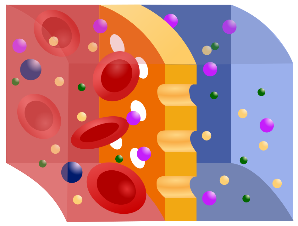
Osmosis involves the special movement of water across a semi-permeable membrane. Water requires a special protein channel in the cell membrane called an aquaporin, which is like a straw that is always open on both sides of the cell membrane. Water also moves from an area of higher concentration to an area of lower concentration down the concentration gradient. When solutes cannot move across the membrane, water moves to try to balance everything out. This, in conjunction with filtration, is always occurring in the kidneys in special units called nephrons.
The processes of diffusion and osmosis are constant, and the goal is homeostasis, or a steady state of balance. When your red blood cells are in an isotonic solution, they are in balance. Most of you will be at least observing, monitoring, possibly starting, or administering a dye or medication through an IV catheter. The most common IV solution is 0.9% NaCl and it is used to treat dehydration. There are other solutions you may learn about in your clinical courses, such as Lactated Ringer's, Dextrose, and others. When you are given 0.9% NaCl solution, it is isotonic to the red blood cells. Imagine if the wrong solution or wrong percentage was given to a patient. For example, if they were administered a 1.5% solution of NaCl solution, the red blood cells would be in a hypotonic solution, meaning there would be too much NaCl inside the cells, so water would rush in to try to balance that out. Their cells would swell up, and could potentially lyse/burst. This is also what happens during a blood transfusion reaction with the wrong blood type, or an infusion that occurs too quickly. It also happens to us naturally when we eat too much salt (a highly salty steak, salty chips, salty movie theater butter popcorn, etc...). It is also the principle behind why we as humans, and even our pets, are unable to swallow salty ocean water without getting sick and nauseous and vomiting. It puts our cells in a hypotonic solution.
On the other hand, if your patient is dehydrated and does not have enough fluid, their red blood cells are in a state of being hypertonic. There is too little NaCl inside the red blood cells, and more outside of them within the extracellular fluids, so the water rushes out to try to balance it out. In the process, the cells crenate (shrink) like a prune. This is why 0.9% NaCl solution is so beneficial to treating dehydration and putting the cells back in an isotonic solution. This occurs naturally when you swim in the salty ocean, a chlorinated swimming pool, or take a hot bath in Epsom salts or solutions containing them. Your fingers and toes crinkle up like prunes.
Another clinical picture of simple diffusion is the example of a negatively charged dye particle binding to the positive charges across the cell membrane and diffusing into it without any help of ATP energy or a protein carrier or channel. Contrast dyes are used in some types of scans, including CT-scans with contrast, MRI with contrast, and PET scans. Kidney pyelograms use this technology to view the kidneys. Angiograms use this technology to look at the arteries and veins of the heart. Barium is used for fecal defagrams, viewing the colon, or looking at the esophagus and stomach.
Osmosis involves the special movement of water across a semi-permeable membrane. Water requires a special protein channel in the cell membrane called an aquaporin, which is like a straw that is always open on both sides of the cell membrane. Water also moves from an area of higher concentration to an area of lower concentration down the concentration gradient. When solutes cannot move across the membrane, water moves to try to balance everything out. This, in conjunction with filtration, is always occurring in the kidneys in special units called nephrons.
The processes of diffusion and osmosis are constant, and the goal is homeostasis, or a steady state of balance. When your red blood cells are in an isotonic solution, they are in balance. Most of you will be at least observing, monitoring, possibly starting, or administering a dye or medication through an IV catheter. The most common IV solution is 0.9% NaCl and it is used to treat dehydration. There are other solutions you may learn about in your clinical courses, such as Lactated Ringer's, Dextrose, and others. When you are given 0.9% NaCl solution, it is isotonic to the red blood cells. Imagine if the wrong solution or wrong percentage was given to a patient. For example, if they were administered a 1.5% solution of NaCl solution, the red blood cells would be in a hypotonic solution, meaning there would be too much NaCl inside the cells, so water would rush in to try to balance that out. Their cells would swell up, and could potentially lyse/burst. This is also what happens during a blood transfusion reaction with the wrong blood type, or an infusion that occurs too quickly. It also happens to us naturally when we eat too much salt (a highly salty steak, salty chips, salty movie theater butter popcorn, etc...). It is also the principle behind why we as humans, and even our pets, are unable to swallow salty ocean water without getting sick and nauseous and vomiting. It puts our cells in a hypotonic solution.
On the other hand, if your patient is dehydrated and does not have enough fluid, their red blood cells are in a state of being hypertonic. There is too little NaCl inside the red blood cells, and more outside of them within the extracellular fluids, so the water rushes out to try to balance it out. In the process, the cells crenate (shrink) like a prune. This is why 0.9% NaCl solution is so beneficial to treating dehydration and putting the cells back in an isotonic solution. This occurs naturally when you swim in the salty ocean, a chlorinated swimming pool, or take a hot bath in Epsom salts or solutions containing them. Your fingers and toes crinkle up like prunes.
Passive Transport Mechanisms:
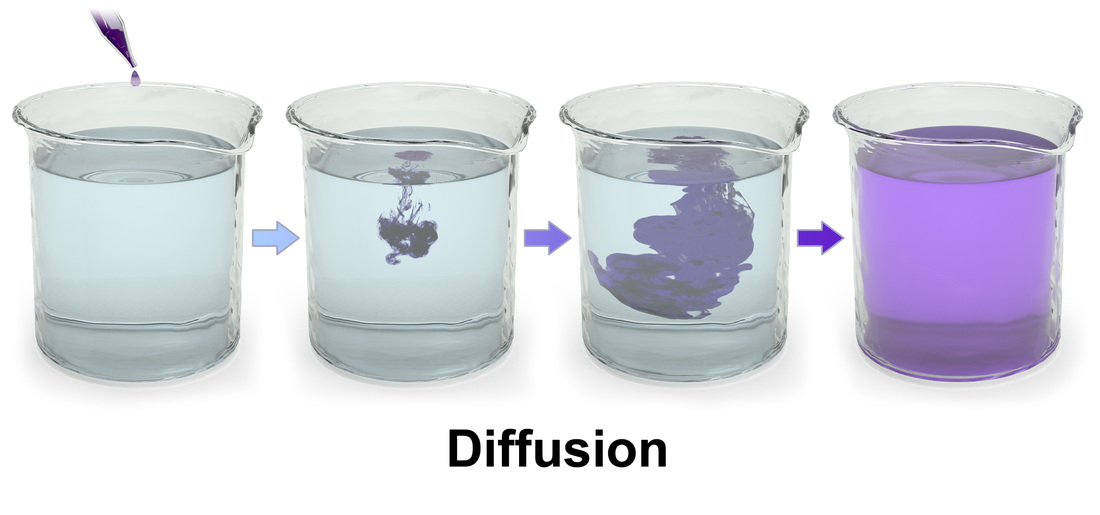
Diffusion:
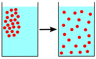
There are several types of diffusion that will be discussed here, but overall, diffusion is based on the principle that molecules have inherent molecular energy (motion) referred to as kinetic energy, in which molecules are in constant motion, which is random. This causes them to bump into each other and collide and change direction. This is sometimes referred to as Brownian Motion. Because some of these particles may be charged, and because differences in concentration may exist, a concentration gradient may be present, where the concentration of solute may be more on one side of a membrane than another. Eventually, molecules will try to reach equilibrium and will become evenly distributed throughout the solution or environment, whether it be a liquid or gas, like air. Some examples include a drop of dye that eventually becomes evenly distributed with water, or room freshener sprayed into the air, which eventually becomes evenly distributed in the air.
Diffusion is basically the net movement of molecules from a region or area of higher concentration to a region or area of lower concentration, moving down the concentration gradient (downhill) driven by kinetic energy produced by the motion of the molecules themselves. The rate of diffusion is affected by various things, including the size and molecular weight of the molecules themselves, their charges, the temperature, and the level of the concentration gradient. If a physical barrier or membrane exists, it will control the diffusion of particles in and out of the cell. Smaller molecules are able to pass through on their own by dissolving in the phospholipids of the membrane or pass through pores in the membrane. If no membrane exists, particles, if soluble, will dissolve in water. This is called simple diffusion and it requires no energy. The special diffusion of water across a semipermeable membrane is referred to as osmosis.
Diffusion is basically the net movement of molecules from a region or area of higher concentration to a region or area of lower concentration, moving down the concentration gradient (downhill) driven by kinetic energy produced by the motion of the molecules themselves. The rate of diffusion is affected by various things, including the size and molecular weight of the molecules themselves, their charges, the temperature, and the level of the concentration gradient. If a physical barrier or membrane exists, it will control the diffusion of particles in and out of the cell. Smaller molecules are able to pass through on their own by dissolving in the phospholipids of the membrane or pass through pores in the membrane. If no membrane exists, particles, if soluble, will dissolve in water. This is called simple diffusion and it requires no energy. The special diffusion of water across a semipermeable membrane is referred to as osmosis.
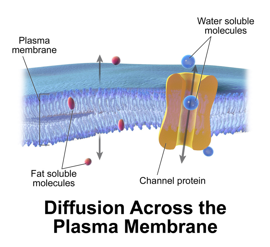
1) Simple Diffusion:
- Higher to lower concentration
- Down the concentration gradient
- No ATP energy required
Smaller or uncharged particles move directly across the phospholipid membrane if they are lipid-soluble. Examples include CO2/O2 exchange in the lungs.
|
Soluble particles dissolving in water
|
Water moving across the semipermeable cell membrane through special pores called aquaporins (osmosis)
|
Simple diffusion of a negatively charged dye particle in water
|
BROWNIAN MOTION AND INHERENT KINETIC ENERGY OF MOLECULES:
|
|
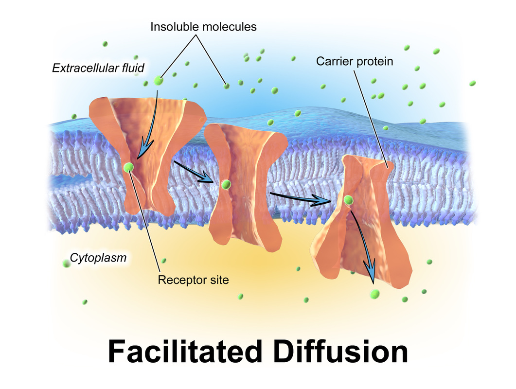
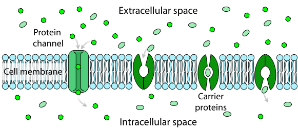
2) Facilitated Diffusion:
Facilitated diffusion still doesn't require energy, however, it does require the the use of some special proteins to "help" substances get across the membrane. These include simple carrier proteins that "squeeze" insoluble molecules across, as well as channel proteins that are always open.
|
|
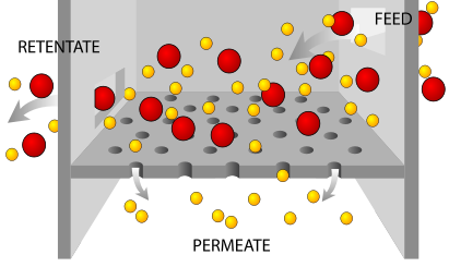
3) Filtration:
In filtration, larger molecules remain trapped on one side of a membrane, whereas smaller molecules go through the tiny pores.
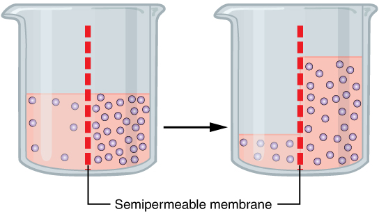
4) Osmosis:
Osmosis is the special diffusion of water across a semipermeable membrane. If there is too much solute, water will rush across the membrane to balance it out and reach equilibrium.
- Isotonic solution: Solution is at equilibrium and there is no net movement of water in or out of the cell; Concentration is the same on both sides of the membrane
- Hypertonic solution: There is a higher concentration of solute outside than inside the fluid of the cell (less inside), so water rushes out of the cell to try to reach equilibrium, and the cell shrinks up/crenates and may die
- Hypotonic solution: There is a lower concentration of solute outside the cell (more inside), so water rushes inside the cell to try to reach equilibrium, and the cell swells up and may burst or lyse
|
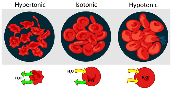
|
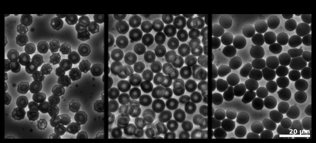
Red blood cells in solutions that are hypertonic, isotonic, and hypotonic to the cells respectively
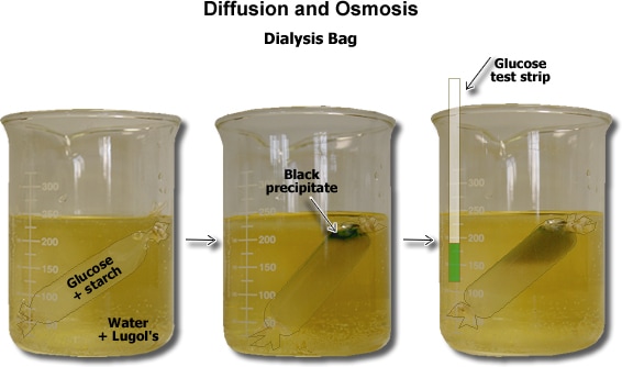
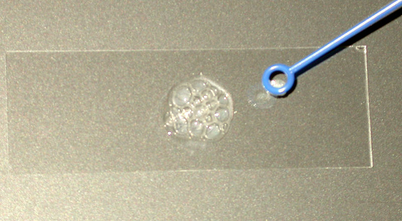
In this experiment, a cell is simulated and placed in a solution by putting starch and glucose solution inside the bag, and iodine solution outside the bag. Iodine complexes with starch and produces a visible color change resulting the lavender-to-purple-to-blue-to-black solution. Do you think the iodine moved into or out of the bag? Hmmmm.....
|
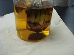
Result of the diffusion-osmosis simulation
|
Active Transport:
In active transport mechanisms, the cell requires ATP energy to move substances across a membrane. These are typically lipid-insoluble molecules, like oil, amino acids, or glucose, and are too large to pass on their own. They need to go through special channels embedded in membrane that require a signal or pump to push them or squeeze them through. They may also be required to move against the concentration gradient or uphill, therefore
- Special carrier proteins may be required (squeeze or thread the molecule through)
- Signals may be required to open up the gated channel protein
- A pump may be required (Na+/K+ pump)
- Require energy
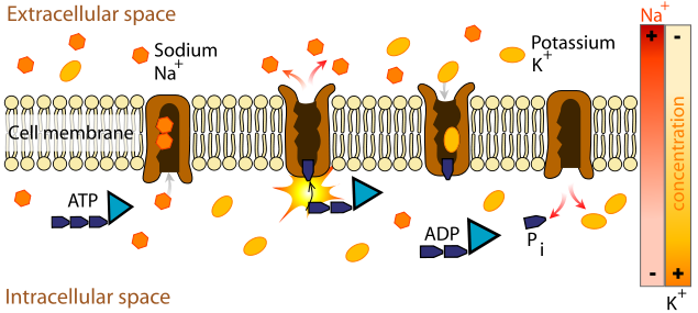
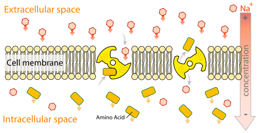
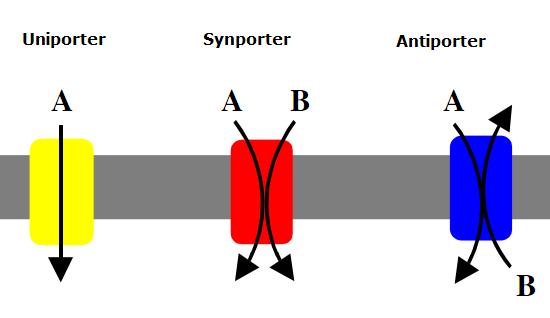
Secondary Active Transport:
These require rotor channels that spin molecules through the membrane, like amino acids or glucose. Molecules may be transported one way, 2 in one direction, or in opposite directions simultaneously.
|
|
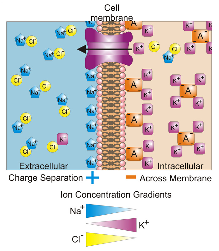
Membrane Potential:
Membrane potential is created across the membrane due to the presence of electrically charged atoms called ions, which create and generate electrochemical potential energy. We know these as "electrolytes", which include things like sodium (Na+), Potassium (K+), Chloride (Cl-), Calcium (Ca2++), Magnesium (Mg+) and others. This is important for muscle movement and function, heart beat, and nerve cell function.
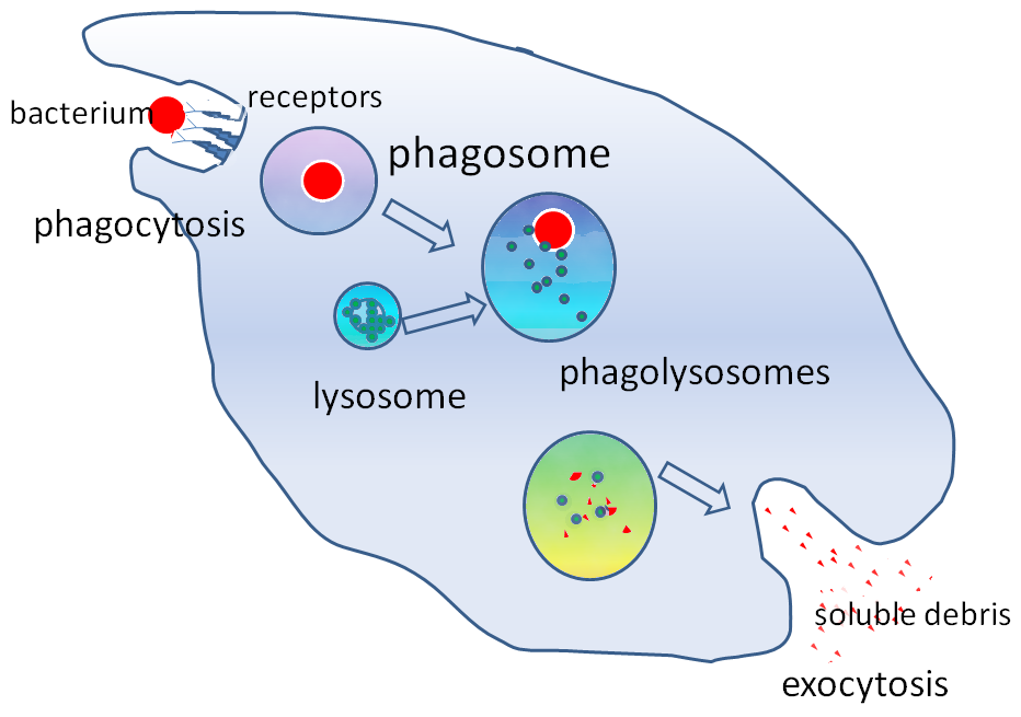
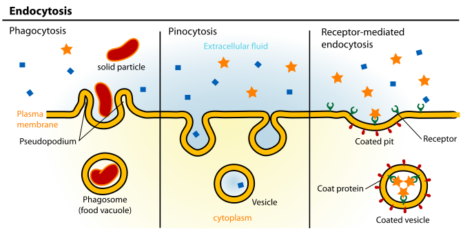
Phagocytosis:
Known as vesicular transport, these mechanisms are forms of active transport that also require energy. This is known as "cellular eating" (phagocytosis) and "cellular drinking" (pinocytosis).
- Endocytosis: particles enter the cell via membrane-bound vesicles
- Exocytosis: particles exit the cell via membrane-bound vesicles

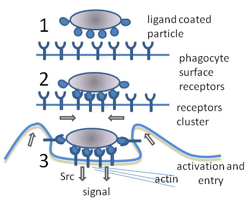
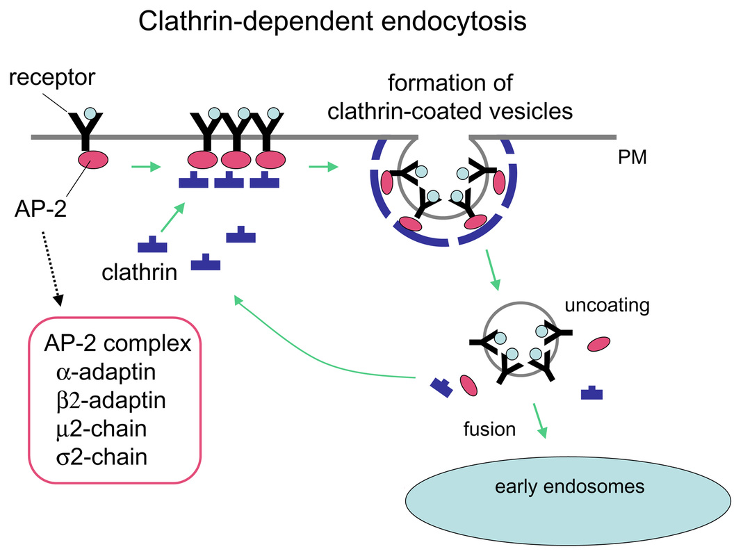
Endocytosis:
In endocytosis, solid or liquid particles enter the cell. Some require receptors for recognition to be let in (receptor-mediated endocytosis).
|

|
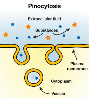
Pinocytosis:
In "cellular drinking", the cell takes in or lets out liquids, such as secretions, mucus, sweat, oil, wax, antibodies, neurotransmitters or other liquid-based substances.
Exocytosis:
In exocytosis, molecules exit the cell in vesicles. Important things include neurotransmitters, hormones, sweat, oil, wax, and mucus.

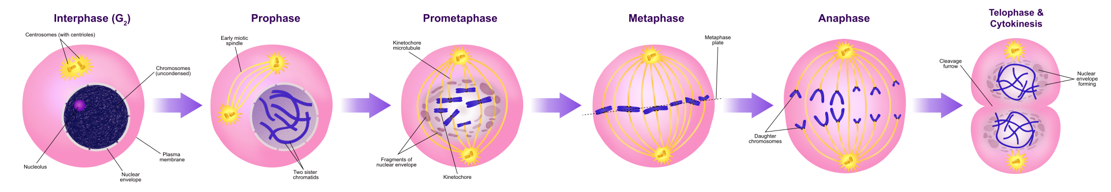
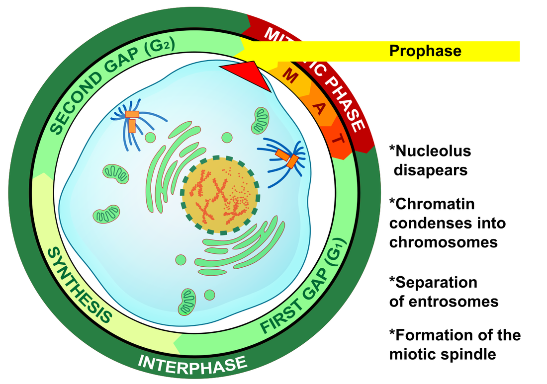
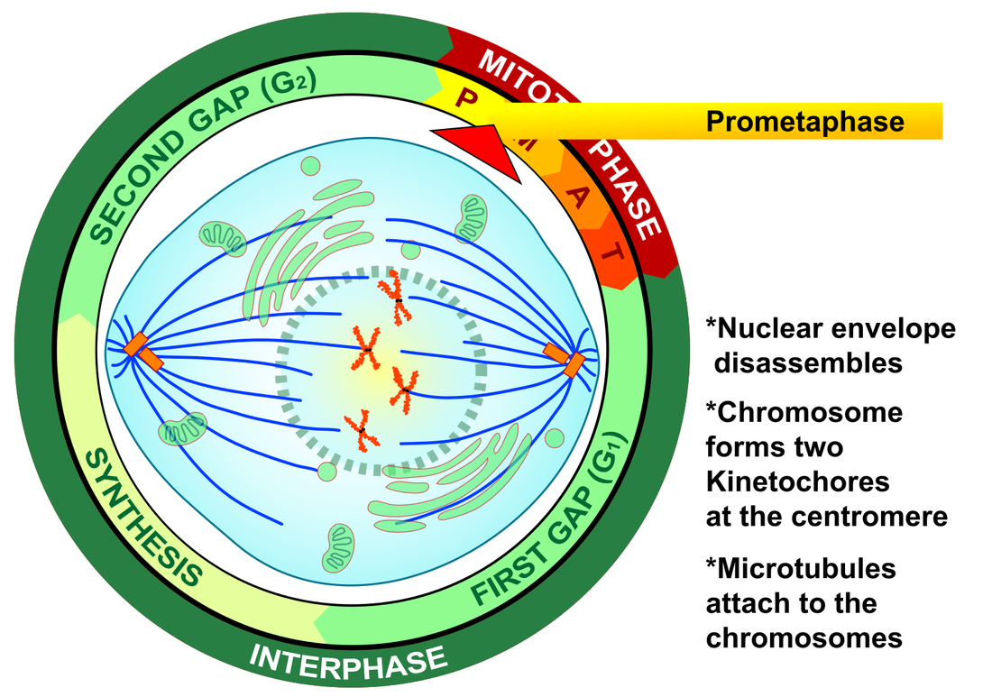
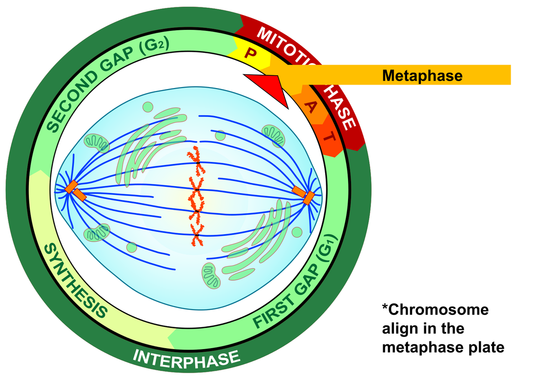
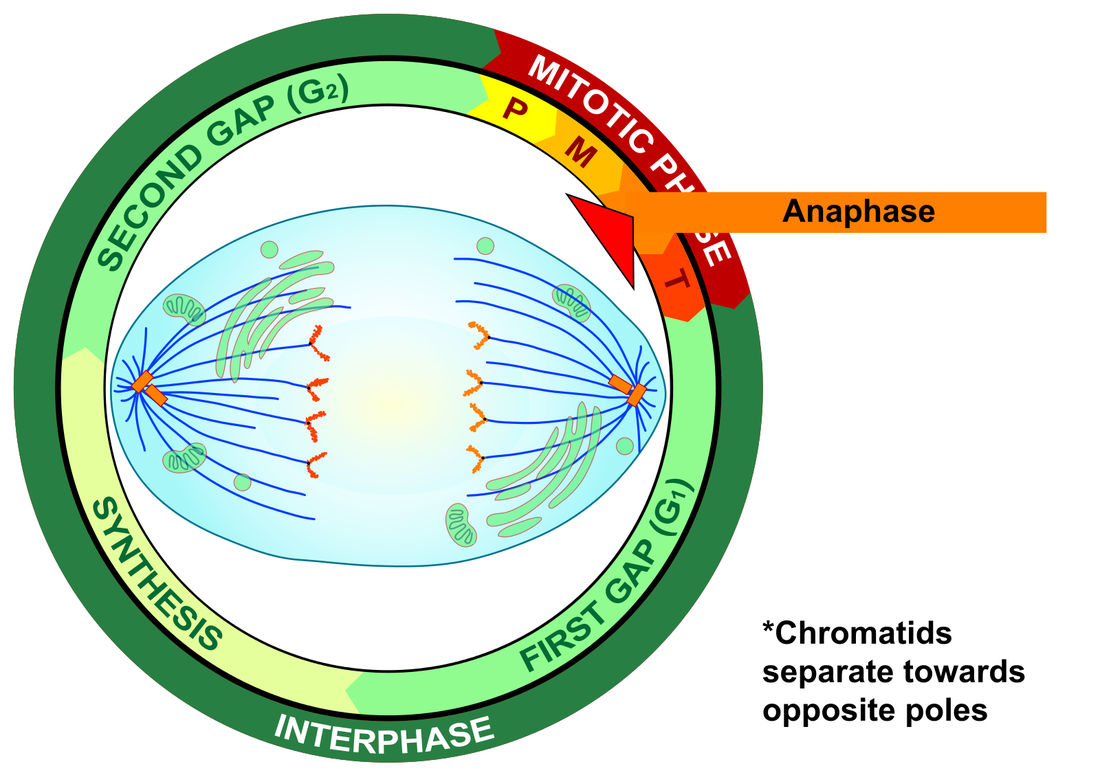
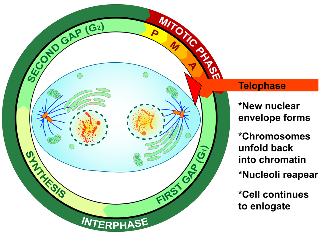
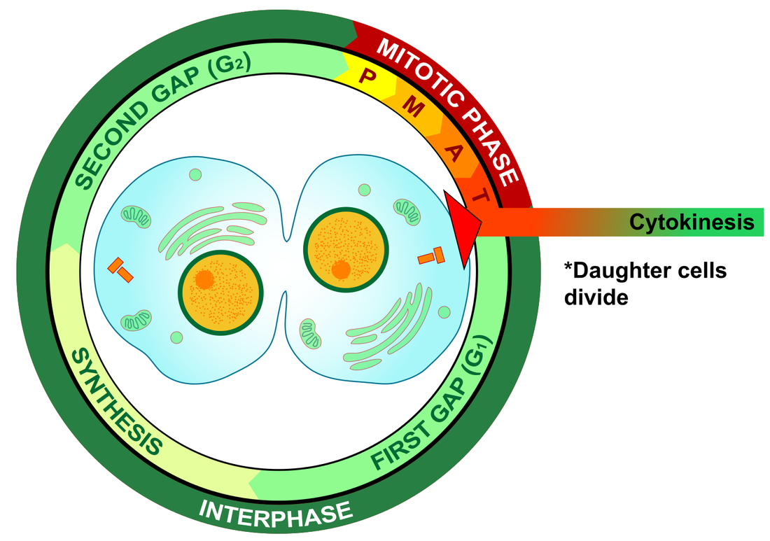
CELL DIVISION:
The Cell Cycle: Mitosis
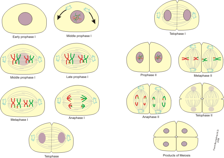
Meiosis: Somatic Cells (Sexual Reproduction: Human Body Cells)
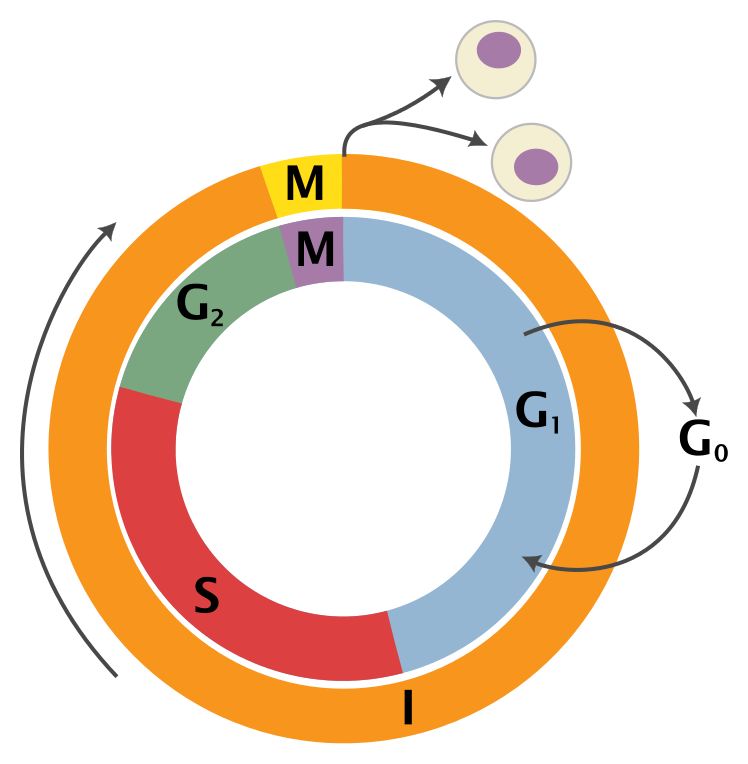
Cell Cycle Regulation:
EXAMPLES OF CELLS UNDER THE MICROSCOPE:
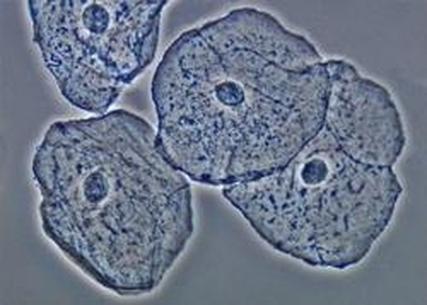
Human cheek cells (squamous epithelial)
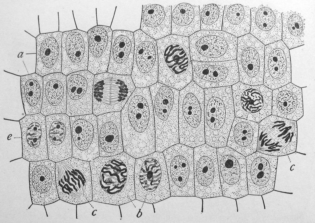
Onion cells

Human cheek cells
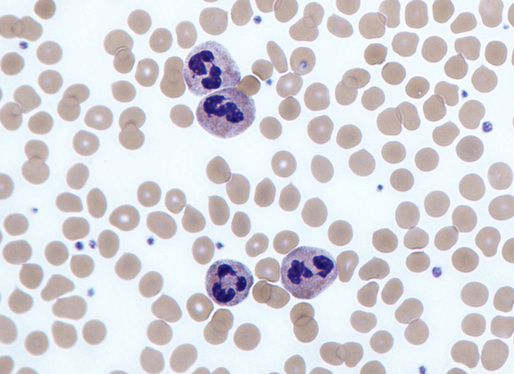
Blood cells: Red blood cells, White blood cells, Platelets
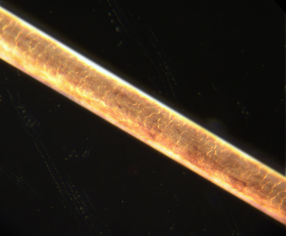
Human Hair
Yeast
|
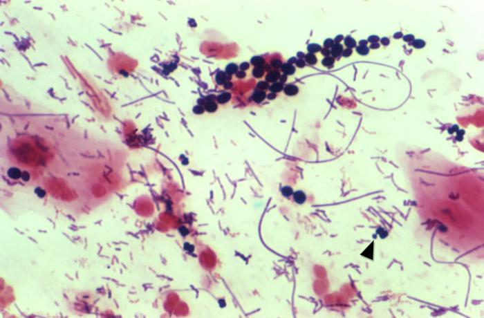
Bacteria
|
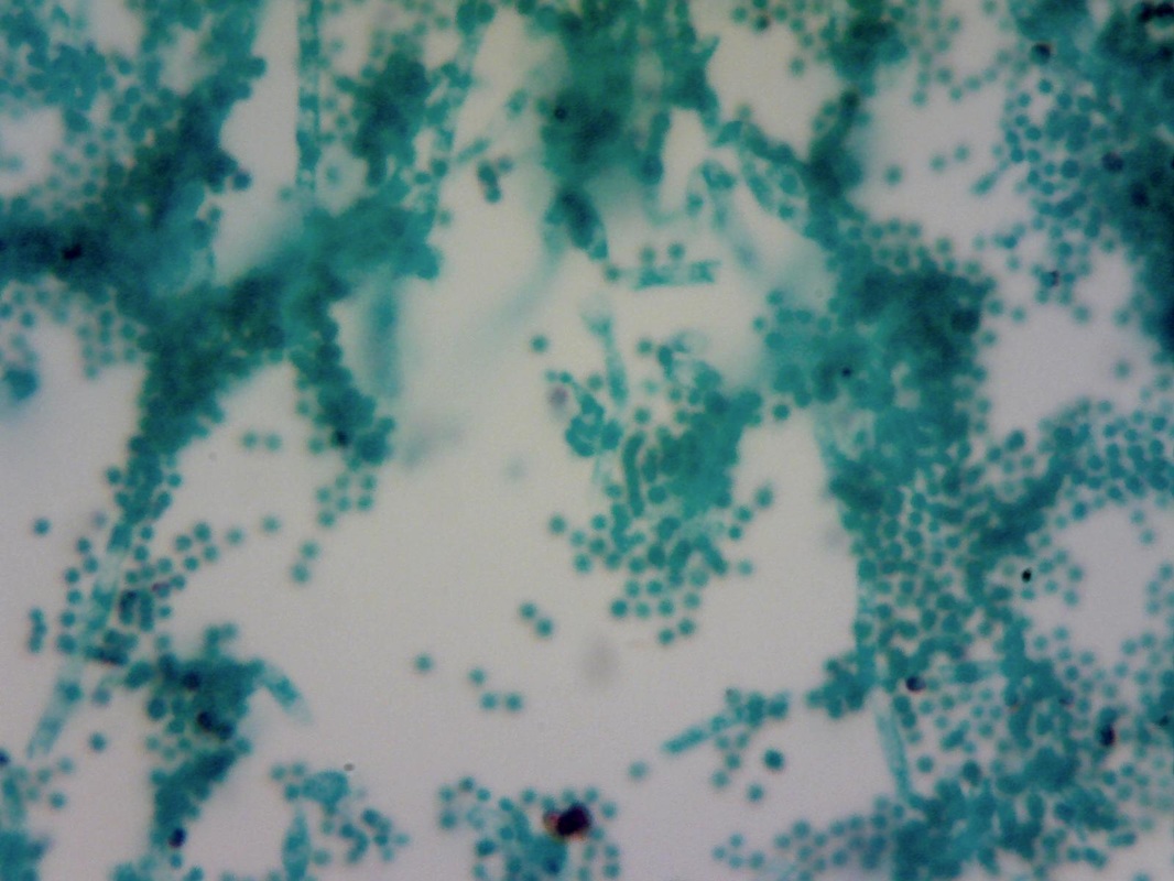
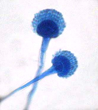
Fungi: Mold
|
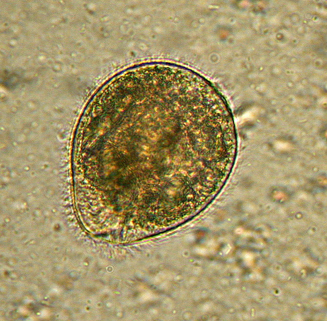
Parasite: Protist
|
Catalase Test: Enzyme breaking apart hydrogen peroxide into oxygen and water
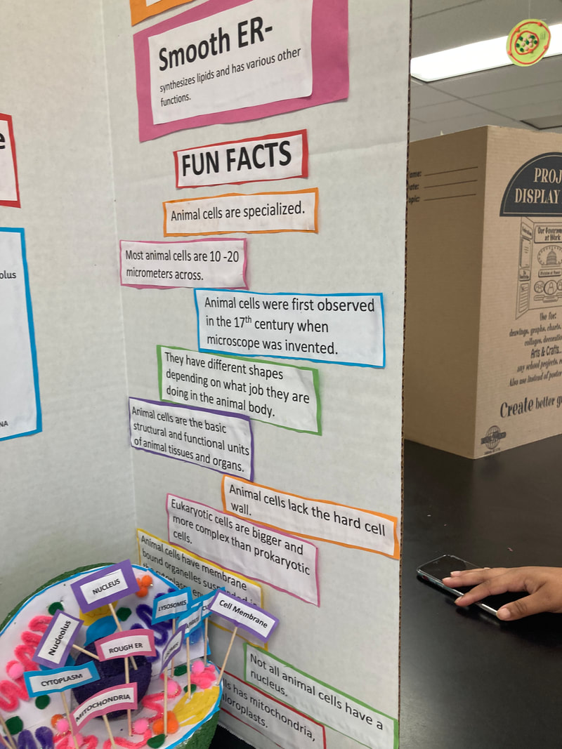
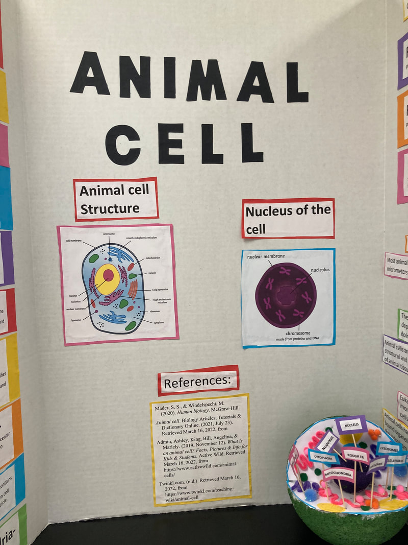
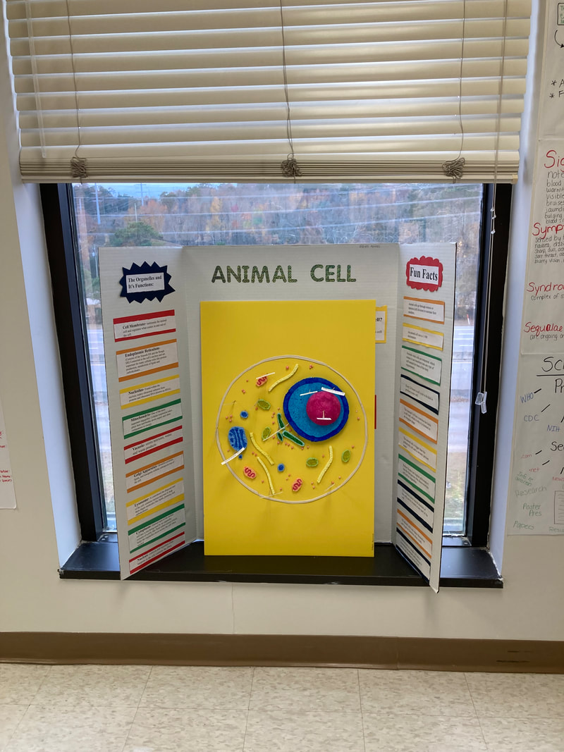
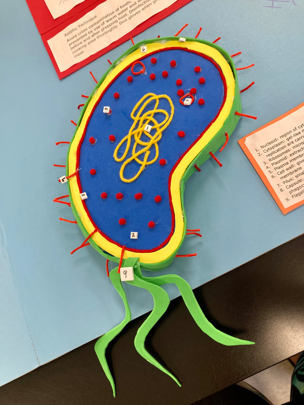
Student Cell Project Examples:
Domain Eukarya (Eukaryote), Kingdom Animalia (Animal):
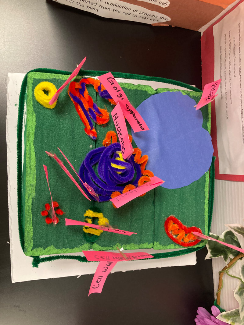
Domain Eukarya (Eukaryote), Kingdom Plantae (Plant):
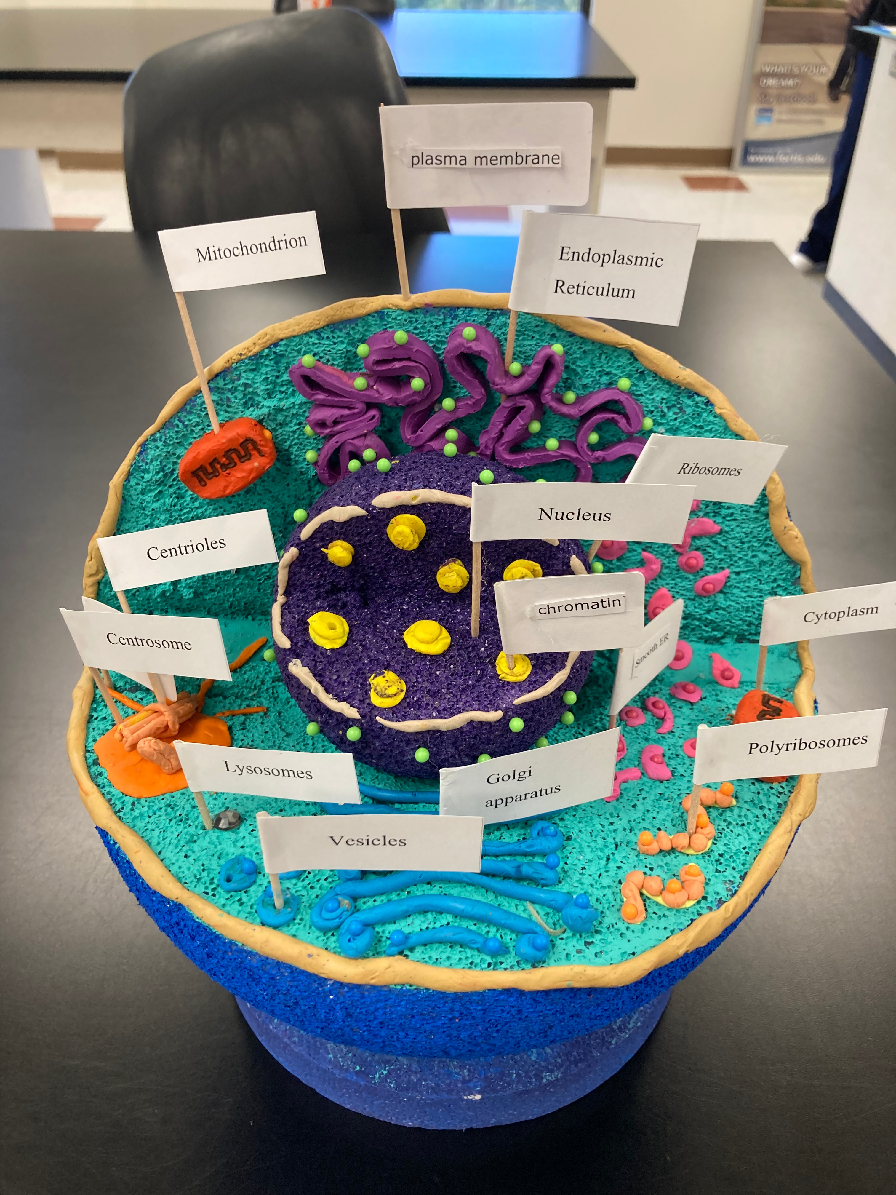
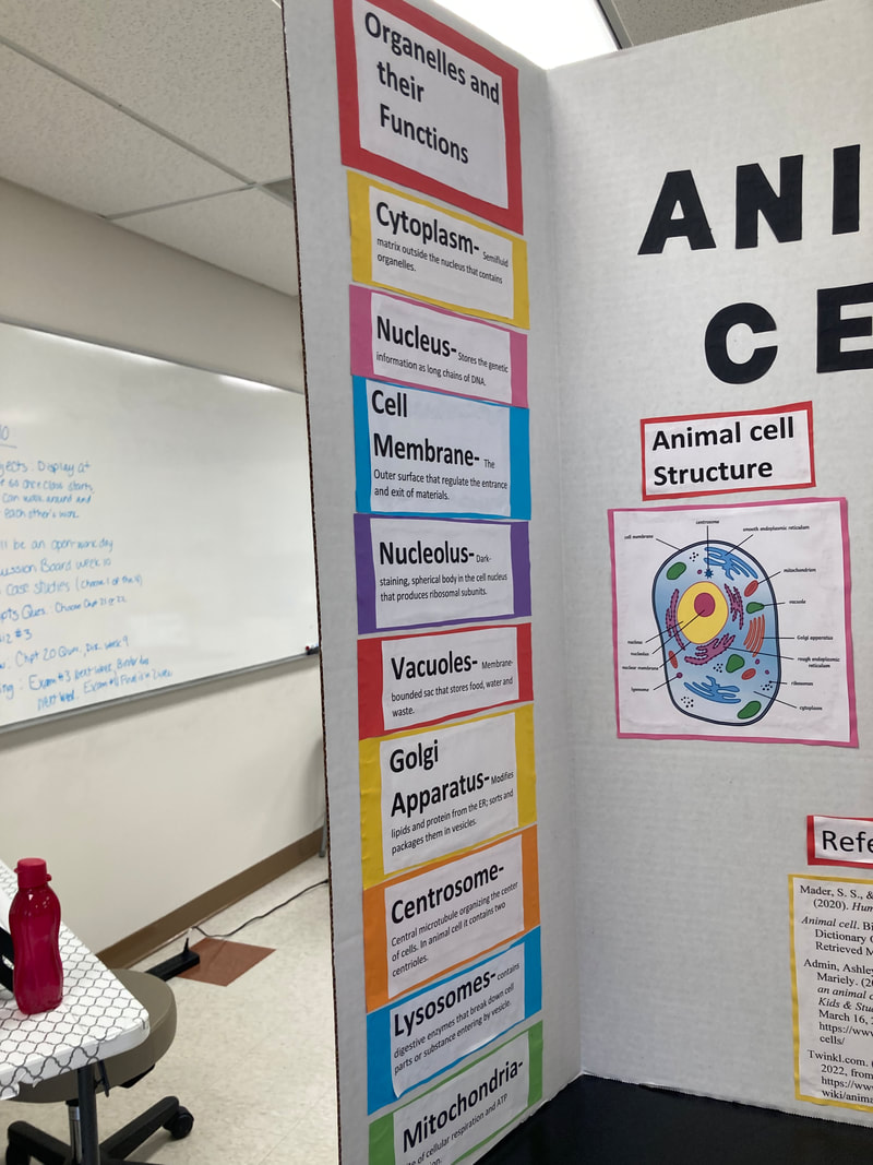
Domain Eukarya (Eukaryote), Kingdom Animalia (Animal Cell):
Domain Bacteria, Kingdom Prokarya (Prokaryote):
Cell Types in the Human Body:
- Red blood cells
- White blood cells
- Platelets
- Skin cells (Squamous Epithelial Cells)
- Epithelial cells
- Endothelial (line organs)
- Goblet (secrete mucous or tears or hormones)
- Ciliated
- Transitional
- Cuboidal
- Endothelial (line organs)
- Cardiomyocytes (heart muscle cells)
- Myocytes (muscle cells)
- Smooth
- Skeletal
- Cardiac (see cardiomyocytes)
- Smooth
- Neurons and neuroglia (brain and spinal cord and nerves)
- Adipocytes (lipids)
- Chondrocytes (cartilage)
- Kupffer cells and hepatocytes (liver)
- Ova
- Sperm
- Photoreceptors: Rods and cones (eyes; color vision)
- Osteocytes (bone)
- Osteoclasts (break down)
- Osteoblasts (build)
- Osteoclasts (break down)
- Enterocytes (intestines)
Abnormal Cells:
- Sickle cells
- Cancer cells
- Misfolded proteins (prions found in Creutzfeldt-Jakob Disease, or tangled plaques found in dementia, Alzheimer's disease, Lewy-body dementia)
Questions:
- How many cells are in the human body?
- How many different types of cells are there in the human body?
- What roles do these specialized cells play?
- What is the role of cells in tissues?
- Where do cells fall on the levels of organization of life?
- How would you define a cell?
- Can you name the cellular organelles and their functions?
- What is the biggest cell in the human body?
- What is the smallest cell in the human body?
- What is the only flagellated cell in the human body?
- How do cells divide?
- What monitors the process of cell division and what happens when these processes are overrun?
- Where do all cell types originate from?
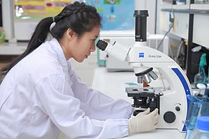
Microscopy:
Objectives:
- Describe and demonstrate how to carry, clean, use and store the compound light microscope.
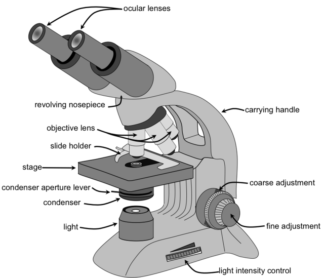
- Identify the parts of the compound light microscope and describe their function.
- Demonstrate how to place a prepared slide on the microscope, and how to properly focus a desired area of a slide under all magnifications.
- Calculate total magnification.
- Lab Manual
- Gateway Virtual Access for Microscopy Lab
- Virtual Slides ("e")
- Virtual Microscope Slides, Coverslips, Etc...
https://upload.wikimedia.org/wikipedia/commons/thumb/1/17/Parts_of_a_Microscope_%28english%29.png/640px-Parts_of_a_Microscope_%28english%29.png
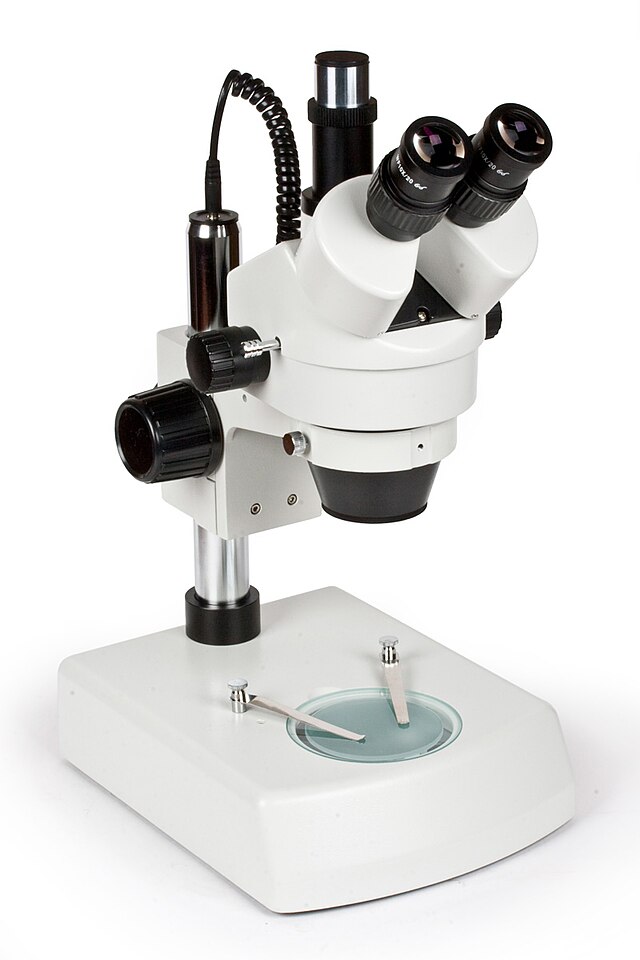
https://upload.wikimedia.org/wikipedia/commons/thumb/5/5d/Microscope_Altami_PS_II.jpg/640px-Microscope_Altami_PS_II.jpg
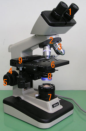
Practice Labeling the Parts of the Microscope:
1.
2.
3.
4.
5.
6.
7.
8.
9.
1.
2.
3.
4.
5.
6.
7.
8.
9.
1) Oculars (eyepieces): 10x magnification (together) - 1-2 removable lenses that you look through to observe the microscopic slide. One of them often has a pointer to identify a particular area on a slide.
2) Revolving Nosepiece: turns and locks into place to change objective lenses.
3) Objective Lenses (Turn, lock into place) - 3-4 on a microscope on the revolving nosepiece
5) Fine focus knob: fine tunes the focus and sharpens the image
6) Stage: flat platform that holds the specimen slide; Stage clips: hold the slide in place; Aperture: the space in the stage that light comes up through from the condenser
7) Light source in the base of the microscope (substage light)
8) Condenser: located just below the stage and moves up and down to adjust the intensity of the light; Iris diaphragm: a lever on the condenser that can be opened/closed to adjust the focus of light (wide/pinpoint)
9) Mechanical stage: knobs that move the stage from side-to-side (bottom) and back-and-forth (top)
Other: light switch (turns the light on/off)
Light intensity knob (dim the light or brighten it)
Arm: back of the microscope that is used to carry/support it from the back
Head: the ocular/eyepieces are attached to the top of the microscope here
Microscope Cleaning: Lens paper and cleaners that are dust-free and optical-safe are used to clean microscope lenses and protect them from being scratched.
Other Terminology:
Field of View - the lighted circular area you see when you bring the microscope into focus; the area of the slide that is being observed; decreases with increasing magnification; increases with decreasing magnification
Magnification
Resolution
Clarity
Parfocal - most microscopes are parfocal and when you move to a different magnification, it is nearly in focus
Stain is applied to cheek cells so that the organelles stand out and can be identified. Cells that line the skin and interior of the mouth fit closely together like floor tile and form a thick layer that protects the underlying tissue from abrasion and microbes. They are superficial and continually slough off and are replaced by underlying cells.
You are able to clearly see the nucleus in the middle, the cytoplasm inside, and the cell membrane of the animal cell.
2) Revolving Nosepiece: turns and locks into place to change objective lenses.
3) Objective Lenses (Turn, lock into place) - 3-4 on a microscope on the revolving nosepiece
- 4x: scanning objective lens - Always begin with this objective lens (shortest barrel; red stripe) to bring your sample into view and focus
- 10x: low-power objective lens
- 40x: high-power objective lens
- 100x: oil-immersion lens
- Total magnification: ocular lenses (10x) x objective lens (example: 4x) = 40x total magnification
5) Fine focus knob: fine tunes the focus and sharpens the image
6) Stage: flat platform that holds the specimen slide; Stage clips: hold the slide in place; Aperture: the space in the stage that light comes up through from the condenser
7) Light source in the base of the microscope (substage light)
8) Condenser: located just below the stage and moves up and down to adjust the intensity of the light; Iris diaphragm: a lever on the condenser that can be opened/closed to adjust the focus of light (wide/pinpoint)
9) Mechanical stage: knobs that move the stage from side-to-side (bottom) and back-and-forth (top)
Other: light switch (turns the light on/off)
Light intensity knob (dim the light or brighten it)
Arm: back of the microscope that is used to carry/support it from the back
Head: the ocular/eyepieces are attached to the top of the microscope here
Microscope Cleaning: Lens paper and cleaners that are dust-free and optical-safe are used to clean microscope lenses and protect them from being scratched.
Other Terminology:
Field of View - the lighted circular area you see when you bring the microscope into focus; the area of the slide that is being observed; decreases with increasing magnification; increases with decreasing magnification
Magnification
Resolution
Clarity
Parfocal - most microscopes are parfocal and when you move to a different magnification, it is nearly in focus
Stain is applied to cheek cells so that the organelles stand out and can be identified. Cells that line the skin and interior of the mouth fit closely together like floor tile and form a thick layer that protects the underlying tissue from abrasion and microbes. They are superficial and continually slough off and are replaced by underlying cells.
You are able to clearly see the nucleus in the middle, the cytoplasm inside, and the cell membrane of the animal cell.
